Intravital Microscopyの技術について
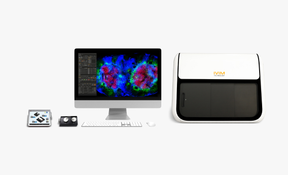
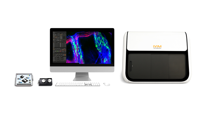
IVIMテクノロジーの革新的なオールインワン生体顕微鏡プラットフォームは、生体内における細胞の複雑かつ動的な挙動を観察できる中核的ソリューションです。この技術は、様々な人間の疾患における根本的な病態生理の解明と、新たな治療法の開発を可能にする最先端のコアテクノロジーです。
Learn more製品紹介
-

IVM-C3
Tractable, Fast, Gentle
IVM-C3は、従来の蛍光顕微鏡と比べて検出効率、光学分解能、画像コントラストを大幅に向上させた高度に統合された共焦点専用生体顕微鏡システムです。
-

IVM-M3
Deep Tissue Imaging, High-Resolution, Tunable
IVM-M3は、波長可変型の柔軟な2光子顕微鏡であり、深部組織の観察に加え、SHG(Second Harmonic Generation)を利用することで、染色を行わずともコラーゲンや線維などの非線形構造を繰り返し観察することが可能なモデルです。
-

IVM-CM3
High Contrast, High Resolution, Dual-mode, Tunable
IVM-CM3は共焦点と2光子の機能を統合したシステムであり、優れたSNR(信号対雑音比)と解像度を提供します。2光子観察のためのフェムト秒レーザーは、690nmから1050nmまでの波長変換が可能です。
-

IVM-MS3
Compact, Cost-Saving, Hands-Free
IVM-MS3は、従来の2光子顕微鏡とは一線を画す、920nm固定式フェムト秒レーザーを搭載したオールインワン2光子生体顕微鏡システムで、非常に簡便かつ高性能です。
-
IVM-CMS3
Cost-Effective, Straightforward, Dual-Mode
IVM-CMS3は、市場で最もコンパクトかつリーズナブルな共焦点・2光子デュアルモードの生体顕微鏡です。簡単なボックス型のオールインワン構造で、生体顕微鏡に必要な機能がほぼすべて搭載されており、当社のベストセラーモデルです。
-

IVM-FS
Practical, Customizable
IVM-FSシステムは、IVM-CMS3と同様の機能を提供しながらも、大型ステージを備えたオープンタイプの生体顕微鏡です。生後6週以上のラットやフェレット、ウサギなどの大型動物にも対応可能です。
-
AI-Image Denoiser
生体内顕微鏡イメージング過程で発生するノイズを効果的に除去するために開発された、自己教師あり学習ベースのアルゴリズムです。
-
IVI Tag™ – In Vivo Labels
生体内の生きた個体において、細胞やタンパク質を卓越した鮮明度で観察します。希釈やシグナルロスを伴うことなく、多色イメージングを実現します。
-
In Vivo Imaging Fixation Adjuncts
動的な臓器を対象に、高精度かつ安定した臓器イメージングを実現します。脳から膵臓まで、動物福祉に配慮した条件下で、生体内研究の可能性を拡張します。
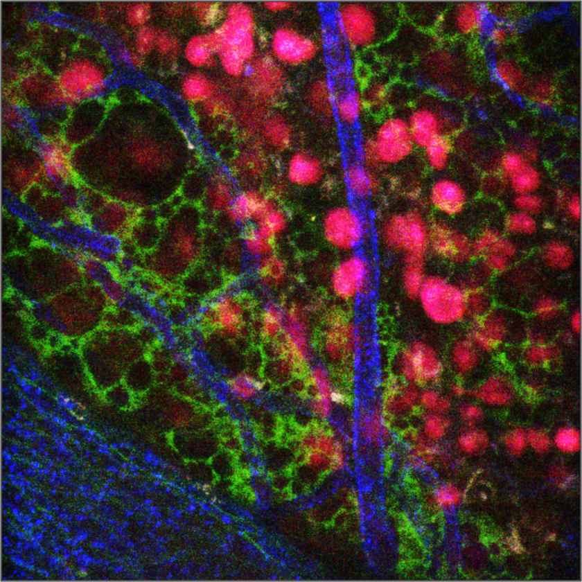
IVM-C3 (Confocal v.3)

IVM-C3 is a highly integrated system for in vivo imaging, offering significantly enhanced detection efficiency, optical resolution, and image contrast compared to conventional fluorescence microscopy. With a 4-wavelength laser and four high-sensitivity confocal detectors, IVM-C3 enables multi-dimensional views of living cells and tissues in 3D or 4D, while supporting up to four different colors.
Key Features
Bone Marrow
Transplanted cell Vessel (CD31)

Bone Marrow
Transplanted cell Vessel (CD31)

Bone Marrow
Transplanted cell Vessel (CD31)

 Shop
Shop Brochures & Flyers
 Contact
IVM-M3 (Two-Photon v. 3)
DeepTissueImaging,High-resolution, Tunable

IVM-M3 seamlessly combines the flexibility of a traditional converted microscope with the high-resolution imaging capabilities of second-harmonic generation microscopy. Featuring a fully automated tunable fs-pulse NIR laser system, IVM-M3 excels in deep tissue imaging by using less-scattering NIR wavelengths. The comprehensive control functionality of the fs- laser system is integrated with the two-photon imaging software, ensuring user convenience with various automation algorithms. Experience advanced imaging with IVM-M3 for your research needs.
Key Features


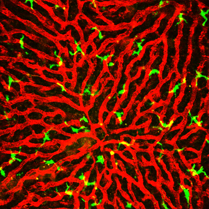
IVM-CM3 (ConfocalandTwo-Photon v. 3)

IVM-CM3 delivers exceptional contrast and resolution with its tunable Two-Photon laser unit.It allows focus on samples at desired wavelengths ranging from as low as 690 nm to as high as 1050 nm, and everything in between. This cutting-edge system seamlessly combines the strengths of both Confocal and Two-Photon microscopy, offering limitless possibilities for three- dimensional imaging of living cells whether near the skin or deep within tumors in small animals. Explore the potential of IVM-CM3 for advanced and versatile imaging in your research.
Key Features
Liver
Fibrotic collagen Hepatocyte

Fibrotic collagen Hepatocyte

Fibrotic collagen Hepatocyte

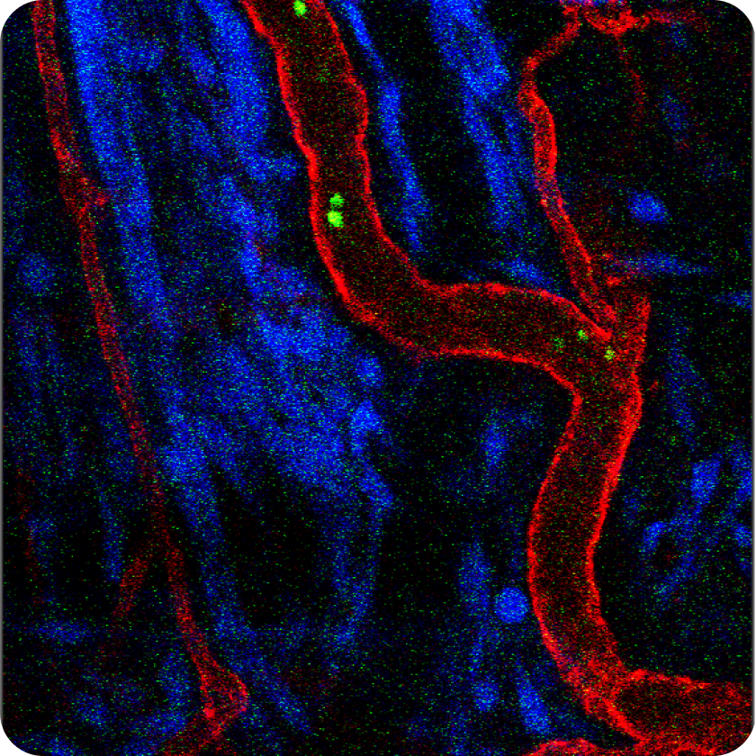
IVM-MS3 (Two- Photon Smart v.3)

IVM-MS3 represents thesmart evolution of IVM-M3, an All-in-One Two-Photon Intravital Microscopy system meticulously optimized for in vivo imaging. This system seamlessly integrates a compact, high-stability, and maintenance-free fs-pulse laser unit into a single box. With the ability to focus on deep tissues at a fixed wavelength of 920nm, IVM- MS3 serves as an exceptional resource for researchers with specific targets, offering optimal functionality within limited resources and budget constraints. Explore the possibilities with IVM-MS3 for efficient and budget-friendly in vivo imaging.
Key Features
Skin
Transplanted cell Transplanted cell Blood vessel (CD31)

Large Intestine
Immune cell Mannose (anti-inflammation) Blood vessel (CD31)

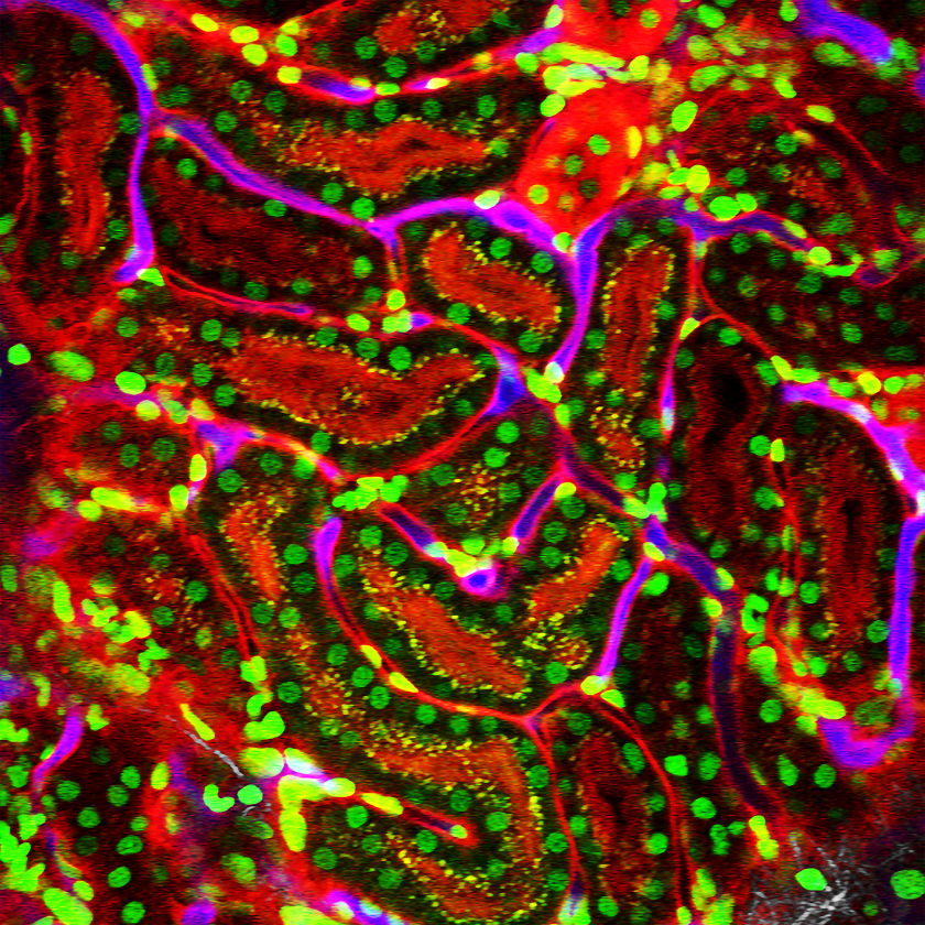
Kidney
H2B (Nuclei) ROSA-mT/mG Blood vessel (CD31)

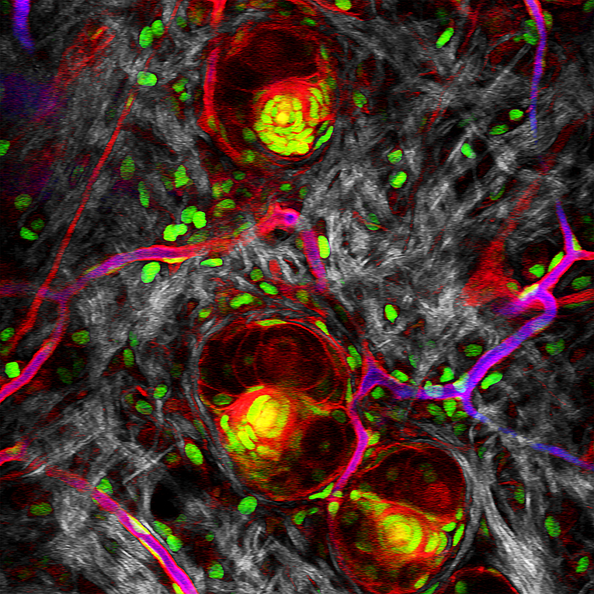
IVM-CMS3 (Confocal and Two-Photon Smart v. 3)

IVM-CMS3 stands out as the world's most compact and affordable dual-mode intravital confocal and two-photon microscope, offering versatile functionality in a single, streamlined box. Leveraging the confocal laser units from IVM-C3 and the compact two-photon laser unit from IVM-MS3, IVM-CMS3 features a one-switch (one-click) mode changing capability, providing convenient multi-purpose use for intravital functional imaging while optimizing space and minimizing costs. Explore the efficient and cost-saving capabilities of IVM-CMS3 for your diverse imaging needs.
Key Features
Skin
CAG-RFP Collagen (SHG)

Liver
Collagen (SHG) Lipid Droplet (SF44)

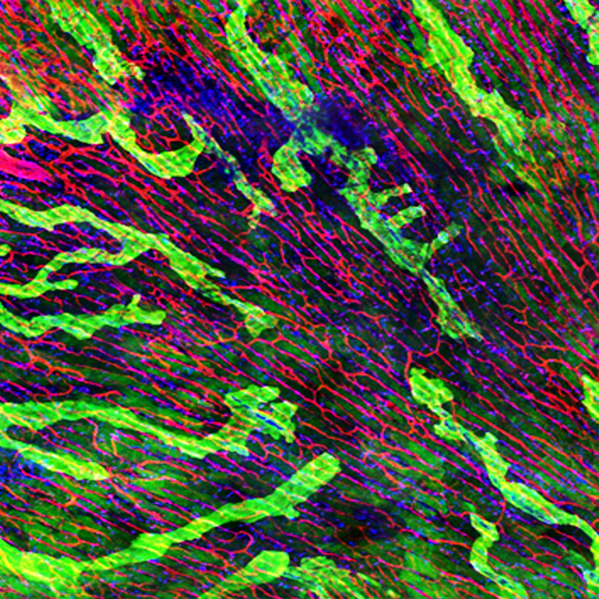
Muscle
Sarcomere (SHG) Nerve (Thy1) Vessel (CD31)

IVM-FS (Free-Space)
Practical, Customizable

The IVM-FS system offers the same functions as the IVM-CMS3 system, with the key difference being its larger stage and modules, designed to accommodate animals larger than mice and rats (older than 6 weeks), such as ferrets, rabbits, and others. With the dark curtain provided, there's no need for a dedicated dark room or specific space just for the microscope.
Key Features
AI-Image Denoiser
A Self-Supervised Learning Algorithm for Reliable Noise Reduction in Intravital Microscopy
The Research Challenge
Intravital microscopy allows researchers to capture dynamic biological events inside living organisms, such as immune cell migration, vascular changes, or rapid drug responses. To observe these processes in real time, imaging must be performed at very high speed.
However, faster imaging inevitably reduces the number of photons collected per frame, resulting in weaker signals and higher noise. This low signal-to-noise ratio (SNR) obscures cell boundaries, blurs fluorescence signals, and introduces uncertainty into quantitative analysis. For researchers, this means unreliable data, reduced reproducibility, and time lost repeating experiments.
The AI-Driven Solution
AI-Image Denoiser addresses these challenges by combining advanced spatio-temporal learning with self-supervised AI algorithms. Unlike conventional AI models that require clean reference images, it learns directly from noisy raw data.
By analyzing spatial and temporal patterns across frames, the software automatically separates true fluorescence signals from random noise, preserving the integrity of biological information.
This means researchers no longer need to prepare special datasets Research Benefits
Transforming Research Efficiency
Conventional denoising can take more than seven hours for a single dataset, slowing down research workflows and limiting the number of experiments that can be conducted.
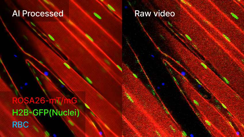
AI-Image Denoiser reduces this process to just 30 minutes, enabling same-day analysis even for complex datasets. Beyond speed, the software preserves fluorescence without distortion, ensuring that fast-moving red blood cells or immune cells can be analyzed accurately. The result is not only higher efficiency but also improved reproducibility—researchers can trust that their quantitative measurements reflect true biological phenomena.
Applications in Advanced Imaging
- Tracking immune cell migration in inflamed tissues
- Monitoring rapid drug responses in vivo
- Measuring blood flow dynamics through red blood cell movement
- Long-term live imaging with reduced photobleaching
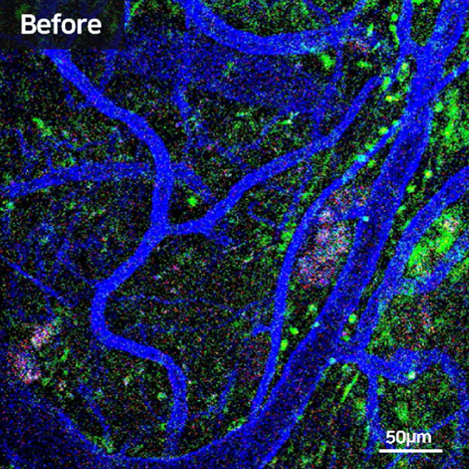
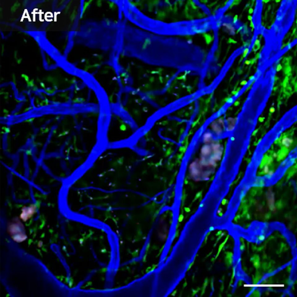
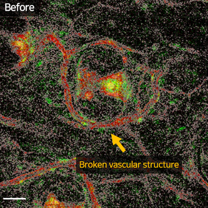
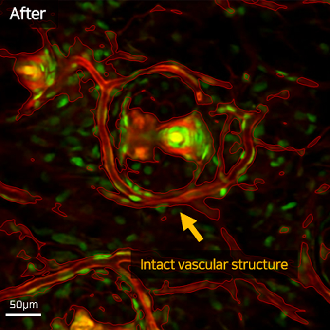
Validation in Real Experiments
In a transgenic mouse ear skin model, intravital microscopy was used to capture vascular structures and circulating red blood cells at high speed. The raw data exhibited strong noise due to limited exposure. After processing with AI-Image Denoiser, the resulting images revealed clear fluorescence signals without distortion and significantly reduced noise. This validation demonstrates that the software provides robust, trustworthy results even under demanding high-speed imaging conditions.
Specifications
| Component | Minimum | Recommended |
|---|---|---|
| GPU | NVIDIA GeForce GTX 1080 | NVIDIA GeForce RTX 4080 |
| RAM | 64 GB | 128 GB |
| CPU | Intel Core i5-11600K, AMD Ryzen 5 3600 | Intel Core i7-13700K, AMD Ryzen 9 5800X |
| Disk | 512 GB SSD (≥150 GB free) | 1 TB NVMe SSD (≥300 GB free) |
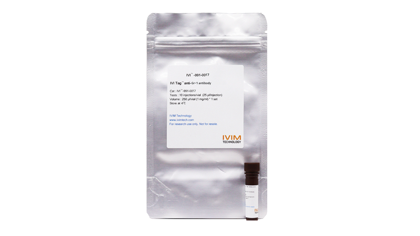
IVI Tag™ – In Vivo Labels
Track cells and proteins inside living organisms with unmatched clarity.
Multi-color imaging, no dilution, no signal loss.
Research Background
Traditional antigen-antibody labeling is limited in vivo due to dilution effects, handling errors, and signal loss. IVIM’s IVI Tag™ overcomes these barriers with advanced fluorescence tagging optimized for intravital microscopy. Researchers can now monitor vascular, immune, and neurological processes in real time without compromising signal integrity.
Core Technologies
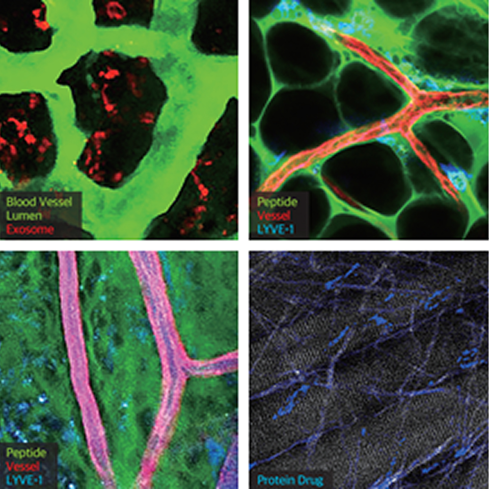
- Direct In Vivo Labeling – No dilution, no handling error, no fluorescence loss
- Multi-Color Compatibility – Four excitation/emission channels + autofluorescence capture (up to 5 signals)
- Organ & Cell-Specific Targets – Endothelial cells (CD31), lymphatic vessels (LYVE-1), dendritic cells (CD11b), T cells (CD3), macrophages (F4/80, CD206), neurons and astrocytes (GFAP, Tau, Amyloid β)
- Broad Microscope Compatibility – Works with IVIM IVM series, confocal, and two-photon systems
- DIY Labeling Kits – Conjugate antibodies, exosomes, peptides, or drugs for customized in vivo imaging
Research Benefits
- Reliable in vivo tracking of immune dynamics, vascular remodeling, and disease progression
- Longitudinal studies without repeated errors from traditional labeling
- High reproducibility and quantitative accuracy for preclinical and neuroscience models
- Easy integration into existing intravital microscopy workflows
Specifications
- Excitation/Emission Wavelengths: 406/445, 495/519, 554/565, 651/667 nm
- Compatibility: IVIM IVM systems, confocal, two-photon microscopes
- Product Range: Antibody panels for CD31, LYVE-1, CD11b, Ly6C, Gr-1, Ly6G, CD3, CD45, F4/80, CD206, GFAP, Tau, Amyloid β, Iba1, and more
DIY In Vivo Labeling Kits
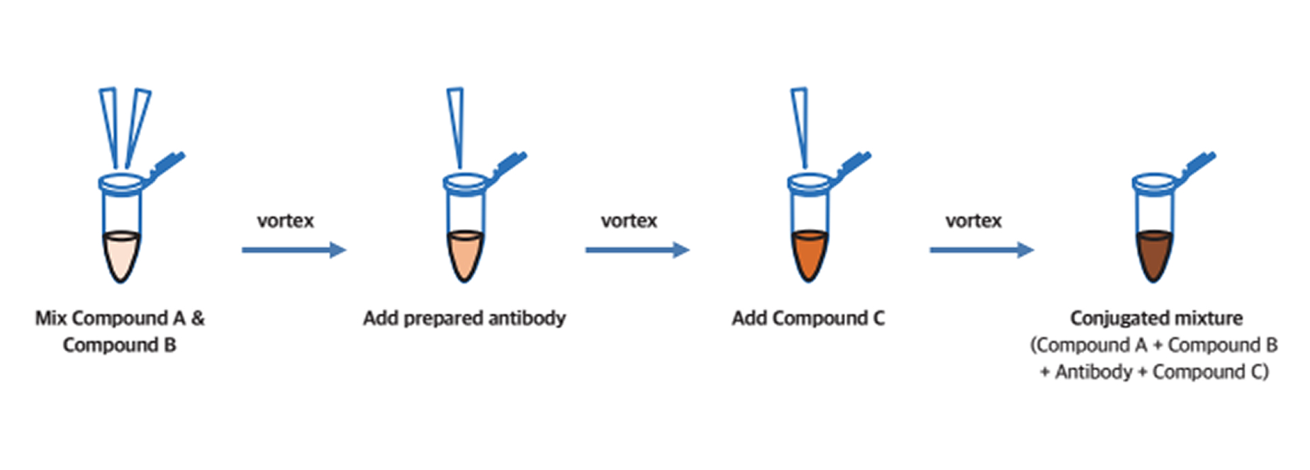
The IVI Tag™ DIY In Vivo Labeling Kit enables researchers to fluorescently tag their own targets—including antibodies, exosomes, peptides, and chemical drugs—for precise in vivo imaging. Using IVIM’s conjugation technology, the kit simplifies the labeling process into a few straightforward steps, making it possible to generate high-quality in vivo labels within hours. Unlike traditional antigen-antibody conjugation methods, IVI Tag™ DIY ensures that fluorophores are expressed at the intended target sites in living tissues, reducing variability and signal loss. This flexibility allows scientists to customize their experiments and track novel biological processes in real time.
In Vivo Label Preparation Steps
Conjugation
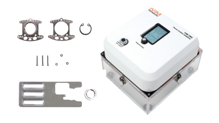
In Vivo Imaging Fixation Adjuncts
Stable, long-term imaging of dynamic organs with precision.
From brain to pancreas, expand research possibilities without compromising animal welfare.
Research Background
Intravital microscopy enables visualization of biological processes in living organisms, but motion artifacts, limited access, and repeated surgeries restrict long-term and reproducible imaging.
Fixation adjuncts solve these issues by stabilizing organs and providing optical access windows, allowing researchers to conduct weeks of continuous observation without sacrificing animals after each session.
Core Technologies
Organ-Specific Windows
Dorsal skinfold, cranial, abdominal, mammary, pancreas, and uterus imaging chambers designed for precise access.
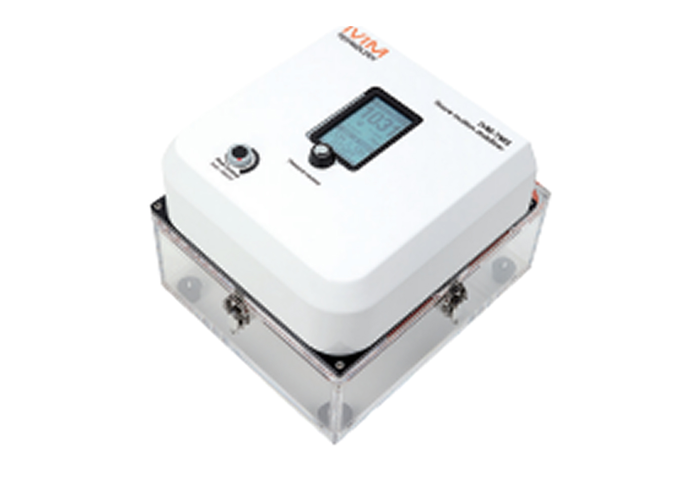
Dynamic Organ Stabilization
Tissue Motion Stabilizer (IVM-TMS) with micro-suction prevents micron-level tissue movement during imaging of the heart, lung, and thymus.
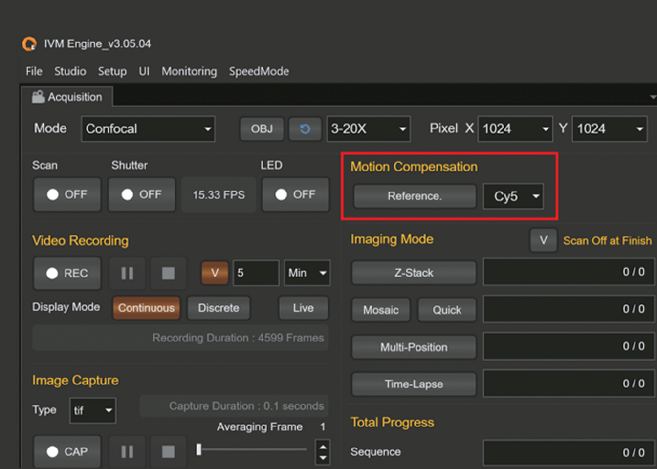
Motion Compensation Software
GPU-assisted algorithm corrects motion artifacts in real time, ensuring high-quality imaging of moving tissues.
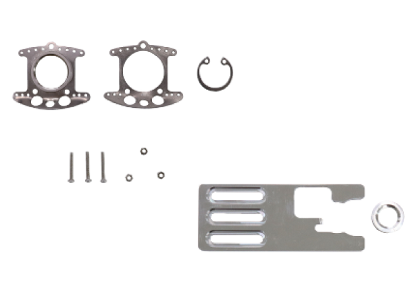
Minimally Invasive Design
Lightweight, compact chambers reduce surgical burden and extend longitudinal studies.
Research Benefits
- Enables longitudinal imaging for several weeks
- Reduces animal use, promoting ethical research practices
- Provides stable platforms for drug delivery, cancer progression, immune dynamics, and vascular studies
- Enhances reproducibility and quantitative reliability in preclinical models
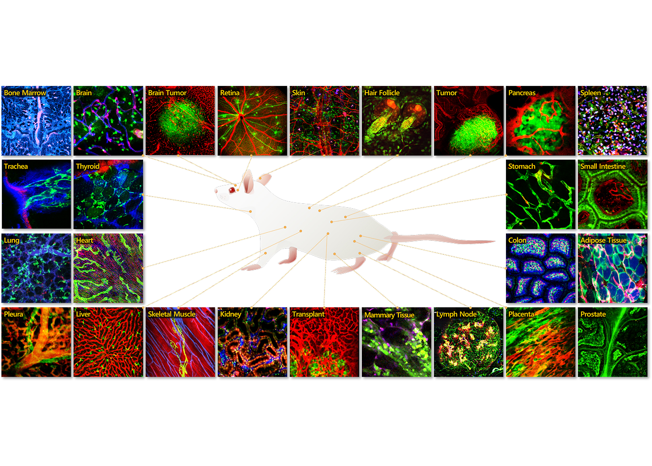
Applications Gallery
-
Lung
Intravital in vivo applications in the lungs of mice provide a powerful tool for studying pulmonary biology, disease processes.
-
Heart
In vivo intravital imaging of the heart in mice offers a powerful approach to studying dynamic cardiac processes and cardiovascular function.
-
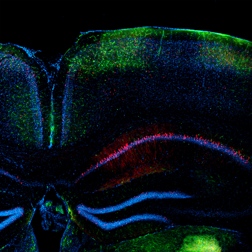
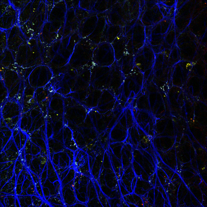
Brain
IVIM Technology provides advanced imaging solutions that support a wide range of intravital imaging applications in the brain.
-
Kidney
In vivo intravital imaging of the kidney offers a powerful approach to studying dynamic cellular and molecular processes within this vital organ.
-
Pancreas
Intravital in vivo applications in the pancreas offer a unique window into the dynamic processes underlying pancreatic physiology.
-
Liver
In vivo intravital imaging of the liver offers a powerful approach to studying dynamic cellular and molecular processes within this vital organ.
-
Tumor
In vivo intravital imaging of the tumor offers you the opportunity to explore various aspects of tumor biology through intravital imaging.
-
Skin
Leveraging intravital imaging techniques, IVIM Technology empowers researchers to gain valuable insights into the dynamic processes occurring within the skin microenvironment in vivo.
CROサービス
IVIMテクノロジーのCROサービスは、非臨床試験段階においてin vivoイメージングによる検証を通じ、薬剤の分布および有効性に関する直接的なデータを提供します。当社は単なるサービス提供にとどまらず、プロジェクトの成功を支える戦略的パートナーシップを提供し、研究開発の革新を支援します。
Learn more
Events
-
Past Webinar
26 Feb 2026
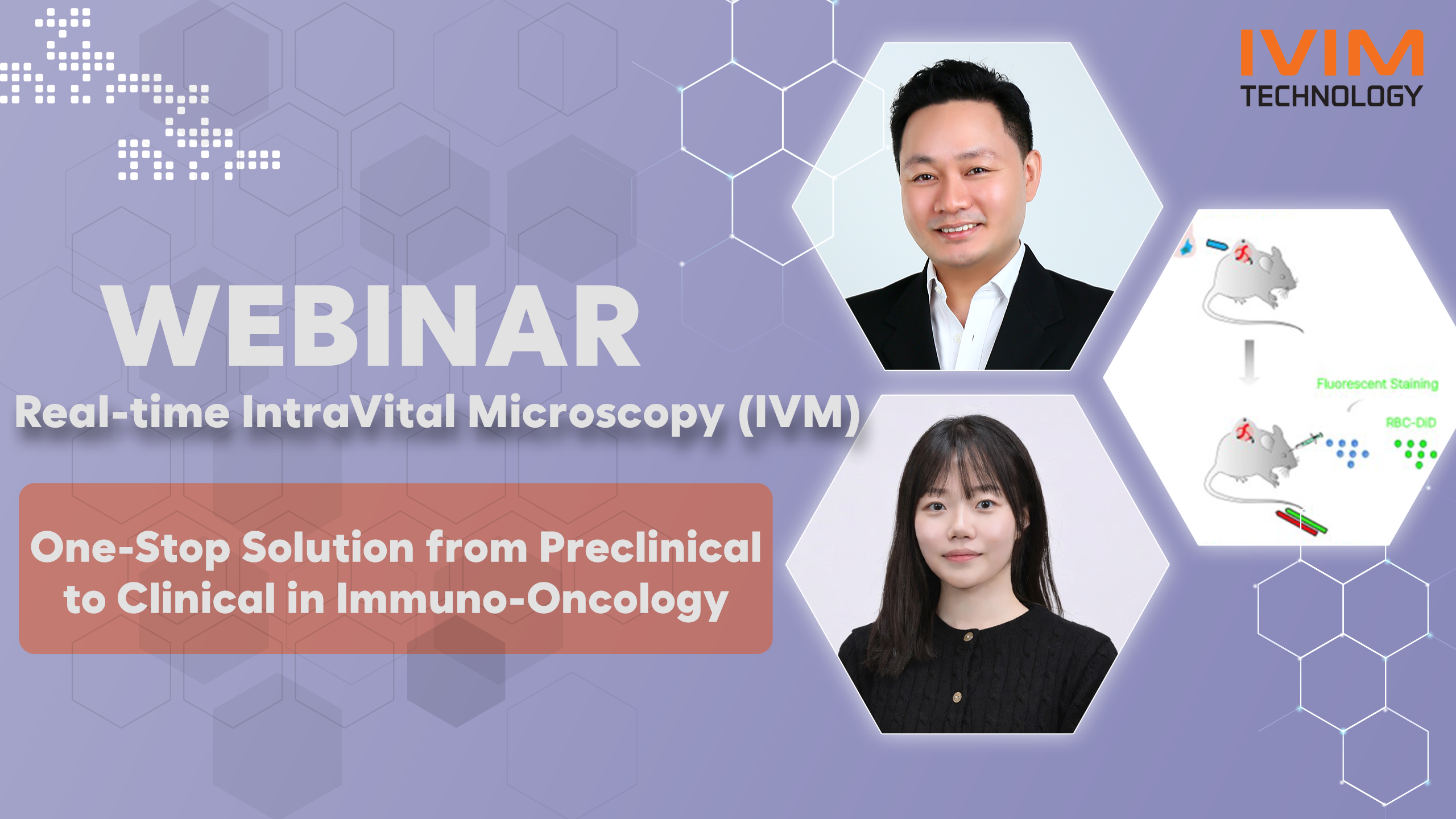
One-Stop Solution from Preclinical to Clinical in Immuno-Oncology
- Jinhyuk Fred CHUNG, PhD
- Chief Science Officer (CSO), INVITROCUE
- Minji GU
- Technical Application Specialist, IVIM Technology Inc.
-
Past Webinar
11 Feb 2026

(EN) Real-time Visualization of In Vivo Immune Responses: A New Paradigm in Preclinical Research
- Pilhan Kim, Ph.D.
- CEO, IVIM Technology Inc.
- Keehoon Jung, Ph.D.
- Associate Professor, Department of Anatomy, Seoul National University College of Medicine
-
Past Webinar
21 Jan 2026

(JP) 腫瘍微小環境ネットワークを標的とした新規治療戦略の開発と可視化
- Pilhan Kim, Ph.D.
- CEO, IVIM Technology Inc.
- Tetsuro Watabe, Ph.D.
- Professor Department of Biochemistry, Graduate School of Medical and Dental Sciences, Institute of Science Tokyo
-
Workshop
05 Mar 2026

2026 Special Event: in vivo Heart Imaging Virtual Demo
Learn more -
Workshop
24 Feb 2026

IVM Imaging Seminar and Workshop
Learn more -
Workshop
12 Jan 2026
LFD/AIM Workshop 2026: FLuorescence Advanced Imaging Research (FLAIR)
Learn more -
Exhibition
26 Jan 2026 - 27 Jan 2026

REGENAGE, Singapore 2026: International Meeting on Ageing and Regenerative Medicine
Learn more -
Exhibition
06 Dec 2025 - 10 Dec 2025
CELL BIO 2025: ASCB–EMBO Joint Meeting
Learn more -
Exhibition
05 Nov 2025 - 09 Nov 2025
SITC 2025: 40th Society for Immunotherapy of Cancer
Learn more
-
Past Webinar
26 Feb 2026
One-Stop Solution from Preclinical to Clinical in Immuno-Oncology
- Jinhyuk Fred CHUNG, PhD
- Chief Science Officer (CSO), INVITROCUE
- Minji GU
- Technical Application Specialist, IVIM Technology Inc.
-
Past Webinar
11 Feb 2026
(EN) Real-time Visualization of In Vivo Immune Responses: A New Paradigm in Preclinical Research
- Pilhan Kim, Ph.D.
- CEO, IVIM Technology Inc.
- Keehoon Jung, Ph.D.
- Associate Professor, Department of Anatomy, Seoul National University College of Medicine
-
Past Webinar
21 Jan 2026
(JP) 腫瘍微小環境ネットワークを標的とした新規治療戦略の開発と可視化
- Pilhan Kim, Ph.D.
- CEO, IVIM Technology Inc.
- Tetsuro Watabe, Ph.D.
- Professor Department of Biochemistry, Graduate School of Medical and Dental Sciences, Institute of Science Tokyo
-
Workshop
05 Mar 2026
2026 Special Event: in vivo Heart Imaging Virtual Demo
Learn more -
Workshop
24 Feb 2026
IVM Imaging Seminar and Workshop
Learn more -
Workshop
12 Jan 2026
LFD/AIM Workshop 2026: FLuorescence Advanced Imaging Research (FLAIR)
Learn more
-
Exhibition
26 Jan 2026 - 27 Jan 2026
REGENAGE, Singapore 2026: International Meeting on Ageing and Regenerative Medicine
Learn more -
Exhibition
06 Dec 2025 - 10 Dec 2025
CELL BIO 2025: ASCB–EMBO Joint Meeting
Learn more -
Exhibition
05 Nov 2025 - 09 Nov 2025
SITC 2025: 40th Society for Immunotherapy of Cancer
Learn more
