Intravital Microscopy
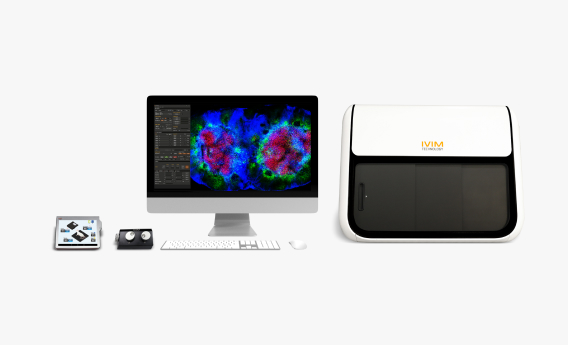
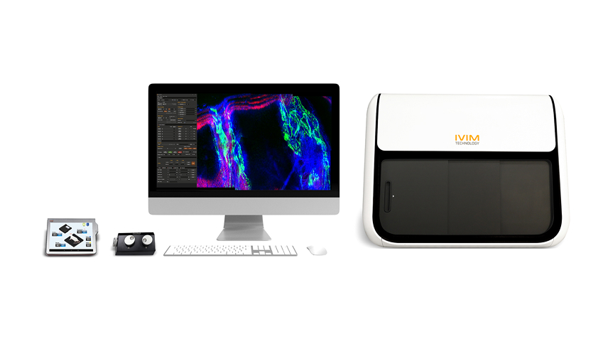
The New All-in-One IVM Series
Confocal and Two-Photon Microscopy System for Live Animal Imaging
Intravital microscopy is a powerful imaging technique that allows real-time visualization of biological processes within living organisms.
Unlike traditional microscopy, which often involves studying fixed or isolated samples, intravital microscopy enables researchers observe dynamic events in their natural physiological context.
This technique is commonly employed in preclinical research in various fields including immunology, neuroscience, and cancer research, thus providing crucial insights into cellular behavior, interactions, and responses within living systems.
Intravital Microscopy Series 3
Have the opportunity to experience the world’s first All-in-One Intravital Microscopy Platform, Series 3, from IVIM Technology
The "New All-in-One IVM Series" stands as a game-changer addressing the critical need for effective cellular validation, tracking, and mode of action determination in real-time. It not only elevates academic research capabilities by providing cutting-edge tools for cellular exploration but also revolutionizes industrial research by expediting drug development timelines and improving overall success rates in the competitive pharmaceutical landscape.
 Shop
Shop Brochures & Flyers
 Contact
Features
Products
-

IVM-C3
Tractable, Fast, Gentle
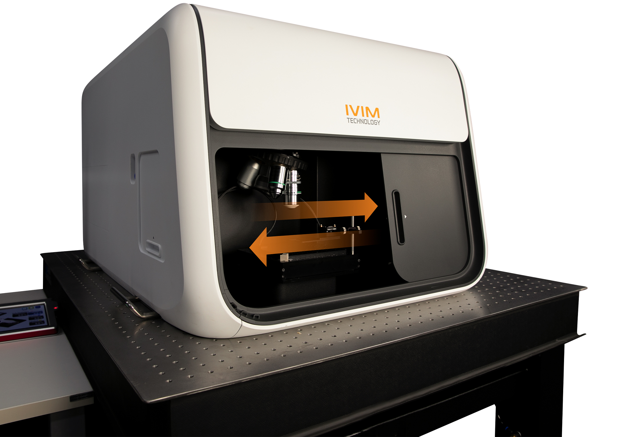
IVM-C3 is a highly integrated system for in vivo imaging, offering significantly enhanced detection efficiency, optical resolution, and image contrast compared to conventional fluorescence microscopy.
-

IVM-M3
Deep Tissue Imaging, High-Resolution, Tunable
IVM-M3 seamlessly combines the flexibility of a traditional converted microscope with the high-resolution imaging capabilities of second-harmonic generation microscopy.
-

IVM-CM3
High Contrast, High Resolution, Dual-mode, Tunable
IVM-CM3 delivers exceptional contrast and resolution with its tunable Two-Photon laser unit, allowing focus on desired wavelengths ranging from as low as 690 nm to higher up to 1050 nm, and everything in between.
-

IVM-MS3
Compact, Cost-Saving, Hands-Free
IVM-MS3 represents the smart evolution of IVM-M3, an All-in-One Two-Photon Intravital Microscopy system meticulously optimized for in vivo imaging.
-
IVM-CMS3
Cost-Effective, Straightforward, Dual-Mode
IVM-CMS3 stands out as the world's most compact and affordable dual-mode intravital confocal and two-photon microscope, offering versatile functionality in a single, streamlined box.
-

IVM-FS
Practical, Customizable
The IVM-FS system offers the same functions as the IVM-CMS3 system, with the key difference being its larger stage and modules, designed to accommodate animals larger than mice and rats (older than 6 weeks), such as ferrets, rabbits, and others. With the dark curtain provided, there's no need for a dedicated dark room or specific space just for the microscope.
-
AI-Image Denoiser
A Self-Supervised Learning Algorithm for Reliable Noise Reduction in Intravital Microscopy.
-
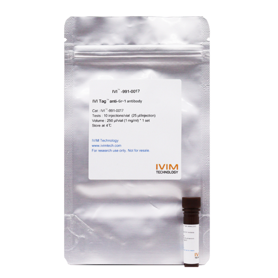
IVI Tag™ – In Vivo Labels
Track cells and proteins inside living organisms with unmatched clarity. Multi-color imaging, no dilution, no signal loss.
-
In Vivo Imaging Fixation Adjuncts
Stable, long-term imaging of dynamic organs with precision. From brain to pancreas, expand research possibilities without compromising animal welfare.
Benefits
IVM-C3 (Confocal v.3)

IVM-C3 is a highly integrated system for in vivo imaging, offering significantly enhanced detection efficiency, optical resolution, and image contrast compared to conventional fluorescence microscopy. With a 4-wavelength laser and four high-sensitivity confocal detectors, IVM-C3 enables multi-dimensional views of living cells and tissues in 3D or 4D, while supporting up to four different colors.
Key Features
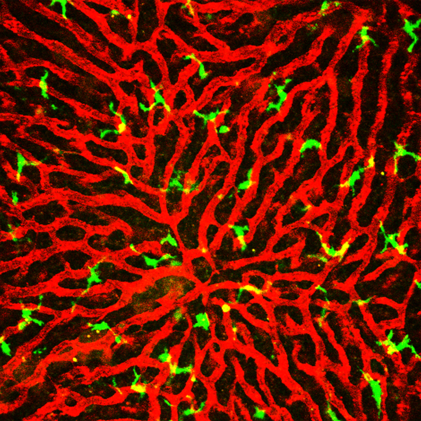
Bone Marrow
Transplanted cell Vessel (CD31)

Bone Marrow
Transplanted cell Vessel (CD31)

Bone Marrow
Transplanted cell Vessel (CD31)

 Shop
Shop Brochures & Flyers
 Contact
IVM-M3 (Two-Photon v. 3)
DeepTissueImaging,High-resolution, Tunable

IVM-M3 seamlessly combines the flexibility of a traditional converted microscope with the high-resolution imaging capabilities of second-harmonic generation microscopy. Featuring a fully automated tunable fs-pulse NIR laser system, IVM-M3 excels in deep tissue imaging by using less-scattering NIR wavelengths. The comprehensive control functionality of the fs- laser system is integrated with the two-photon imaging software, ensuring user convenience with various automation algorithms. Experience advanced imaging with IVM-M3 for your research needs.
Key Features


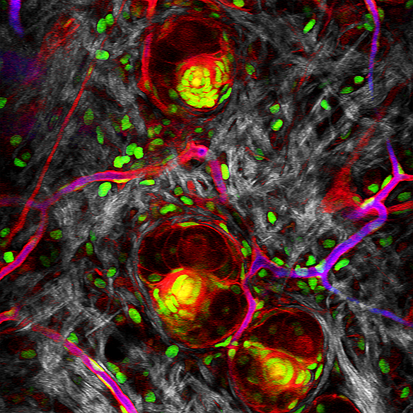
IVM-CM3 (ConfocalandTwo-Photon v. 3)

IVM-CM3 delivers exceptional contrast and resolution with its tunable Two-Photon laser unit.It allows focus on samples at desired wavelengths ranging from as low as 690 nm to as high as 1050 nm, and everything in between. This cutting-edge system seamlessly combines the strengths of both Confocal and Two-Photon microscopy, offering limitless possibilities for three- dimensional imaging of living cells whether near the skin or deep within tumors in small animals. Explore the potential of IVM-CM3 for advanced and versatile imaging in your research.
Key Features
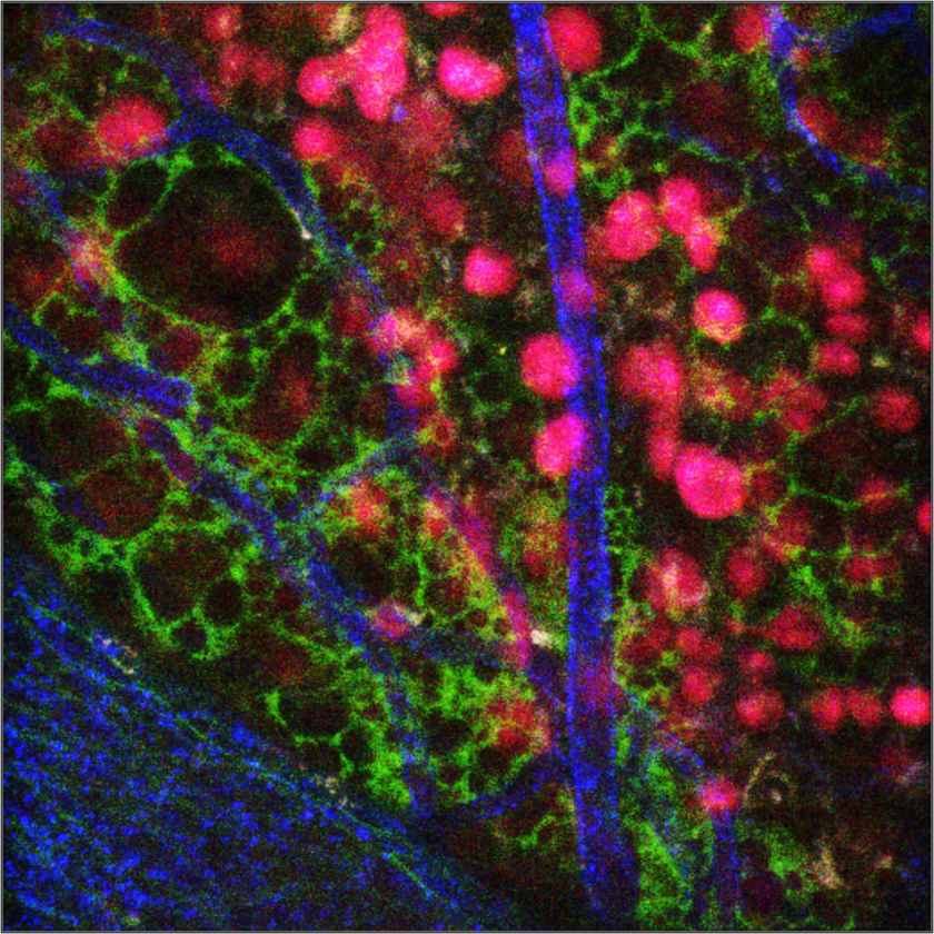
Liver
Fibrotic collagen Hepatocyte

Fibrotic collagen Hepatocyte

Fibrotic collagen Hepatocyte

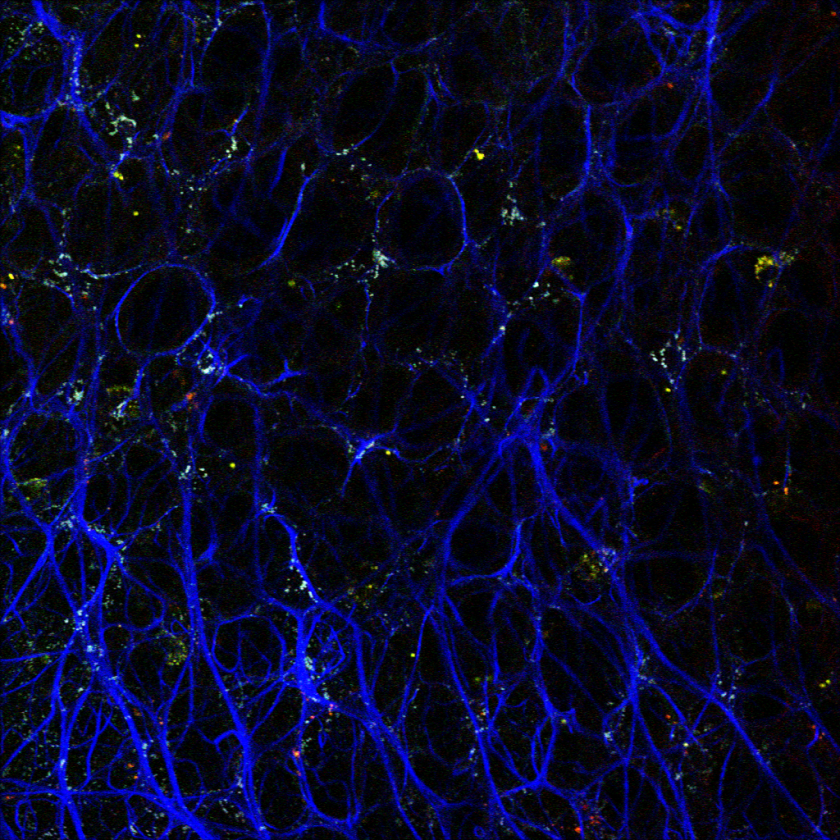
IVM-MS3 (Two- Photon Smart v.3)

IVM-MS3 represents thesmart evolution of IVM-M3, an All-in-One Two-Photon Intravital Microscopy system meticulously optimized for in vivo imaging. This system seamlessly integrates a compact, high-stability, and maintenance-free fs-pulse laser unit into a single box. With the ability to focus on deep tissues at a fixed wavelength of 920nm, IVM- MS3 serves as an exceptional resource for researchers with specific targets, offering optimal functionality within limited resources and budget constraints. Explore the possibilities with IVM-MS3 for efficient and budget-friendly in vivo imaging.
Key Features
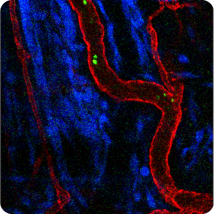
Skin
Transplanted cell Transplanted cell Blood vessel (CD31)

Large Intestine
Immune cell Mannose (anti-inflammation) Blood vessel (CD31)

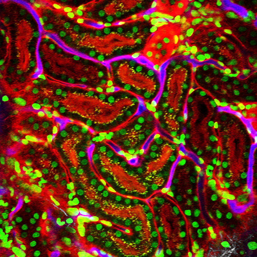
Kidney
H2B (Nuclei) ROSA-mT/mG Blood vessel (CD31)

IVM-CMS3 (Confocal and Two-Photon Smart v. 3)

IVM-CMS3 stands out as the world's most compact and affordable dual-mode intravital confocal and two-photon microscope, offering versatile functionality in a single, streamlined box. Leveraging the confocal laser units from IVM-C3 and the compact two-photon laser unit from IVM-MS3, IVM-CMS3 features a one-switch (one-click) mode changing capability, providing convenient multi-purpose use for intravital functional imaging while optimizing space and minimizing costs. Explore the efficient and cost-saving capabilities of IVM-CMS3 for your diverse imaging needs.
Key Features
Skin
CAG-RFP Collagen (SHG)

Liver
Collagen (SHG) Lipid Droplet (SF44)

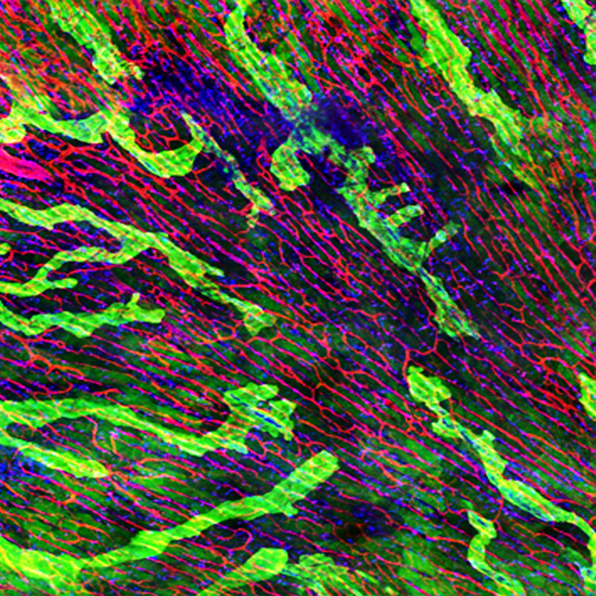
Muscle
Sarcomere (SHG) Nerve (Thy1) Vessel (CD31)

IVM-FS (Free-Space)
Practical, Customizable

The IVM-FS system offers the same functions as the IVM-CMS3 system, with the key difference being its larger stage and modules, designed to accommodate animals larger than mice and rats (older than 6 weeks), such as ferrets, rabbits, and others. With the dark curtain provided, there's no need for a dedicated dark room or specific space just for the microscope.
Key Features
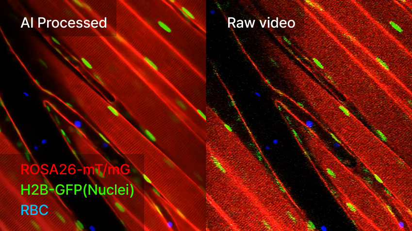
AI-Image Denoiser
A Self-Supervised Learning Algorithm for Reliable Noise Reduction in Intravital Microscopy
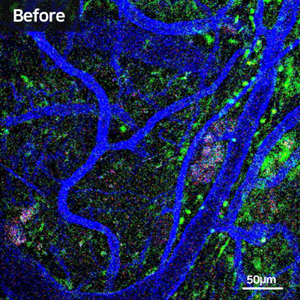
The Research Challenge
Intravital microscopy allows researchers to capture dynamic biological events inside living organisms, such as immune cell migration, vascular changes, or rapid drug responses. To observe these processes in real time, imaging must be performed at very high speed.
However, faster imaging inevitably reduces the number of photons collected per frame, resulting in weaker signals and higher noise. This low signal-to-noise ratio (SNR) obscures cell boundaries, blurs fluorescence signals, and introduces uncertainty into quantitative analysis. For researchers, this means unreliable data, reduced reproducibility, and time lost repeating experiments.
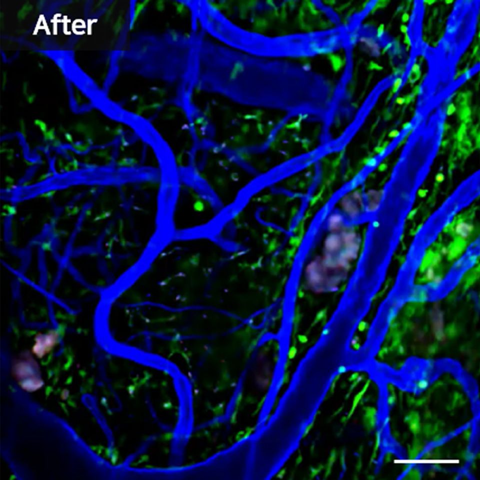
The AI-Driven Solution
AI-Image Denoiser addresses these challenges by combining advanced spatio-temporal learning with self-supervised AI algorithms. Unlike conventional AI models that require clean reference images, it learns directly from noisy raw data.
By analyzing spatial and temporal patterns across frames, the software automatically separates true fluorescence signals from random noise, preserving the integrity of biological information.
This means researchers no longer need to prepare special datasets Research Benefits
Transforming Research Efficiency
Conventional denoising can take more than seven hours for a single dataset, slowing down research workflows and limiting the number of experiments that can be conducted.
AI-Image Denoiser reduces this process to just 30 minutes, enabling same-day analysis even for complex datasets. Beyond speed, the software preserves fluorescence without distortion, ensuring that fast-moving red blood cells or immune cells can be analyzed accurately. The result is not only higher efficiency but also improved reproducibility—researchers can trust that their quantitative measurements reflect true biological phenomena.
Applications in Advanced Imaging
- Tracking immune cell migration in inflamed tissues
- Monitoring rapid drug responses in vivo
- Measuring blood flow dynamics through red blood cell movement
- Long-term live imaging with reduced photobleaching
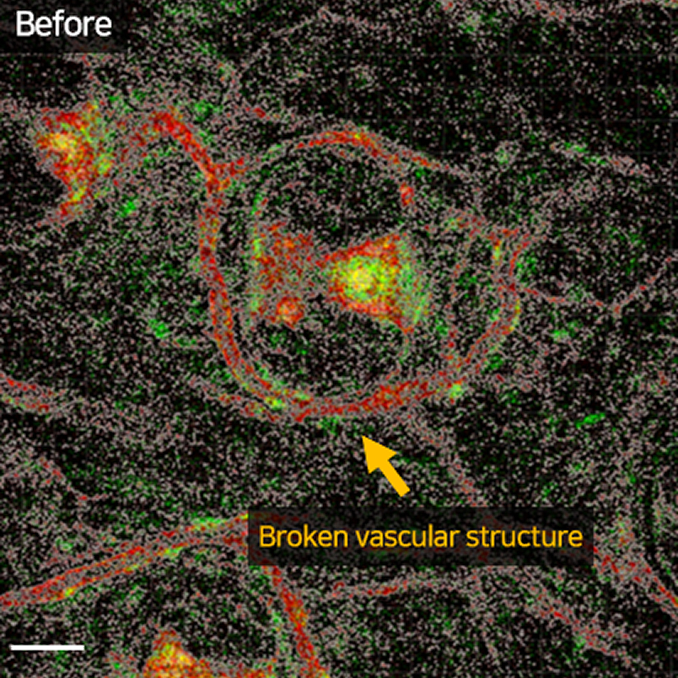
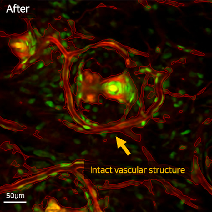
Validation in Real Experiments
In a transgenic mouse ear skin model, intravital microscopy was used to capture vascular structures and circulating red blood cells at high speed. The raw data exhibited strong noise due to limited exposure. After processing with AI-Image Denoiser, the resulting images revealed clear fluorescence signals without distortion and significantly reduced noise. This validation demonstrates that the software provides robust, trustworthy results even under demanding high-speed imaging conditions.
Specifications
| Component | Minimum | Recommended |
|---|---|---|
| GPU | NVIDIA GeForce GTX 1080 | NVIDIA GeForce RTX 4080 |
| RAM | 64 GB | 128 GB |
| CPU | Intel Core i5-11600K, AMD Ryzen 5 3600 | Intel Core i7-13700K, AMD Ryzen 9 5800X |
| Disk | 512 GB SSD (≥150 GB free) | 1 TB NVMe SSD (≥300 GB free) |
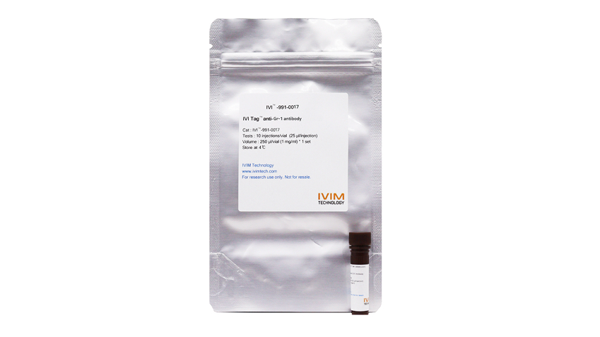
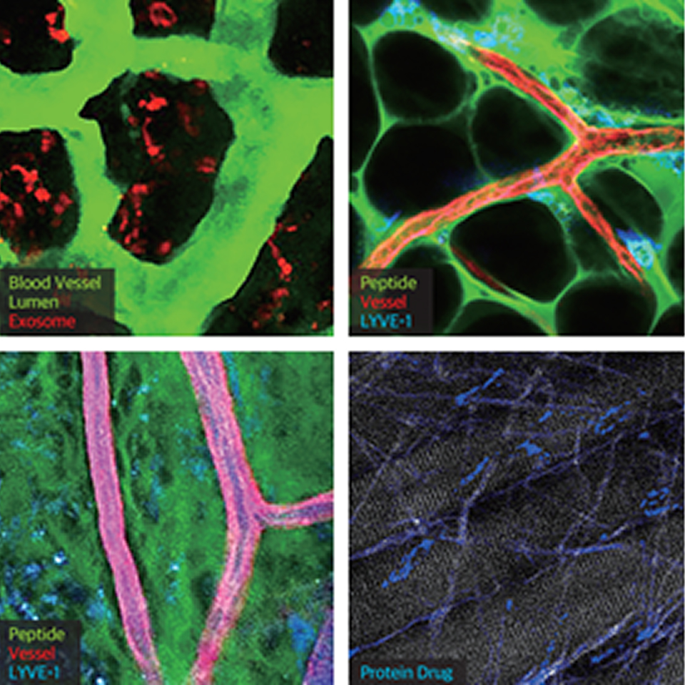
IVI Tag™ – In Vivo Labels
Track cells and proteins inside living organisms with unmatched clarity.
Multi-color imaging, no dilution, no signal loss.
Research Background
Traditional antigen-antibody labeling is limited in vivo due to dilution effects, handling errors, and signal loss. IVIM’s IVI Tag™ overcomes these barriers with advanced fluorescence tagging optimized for intravital microscopy. Researchers can now monitor vascular, immune, and neurological processes in real time without compromising signal integrity.
Core Technologies
- Direct In Vivo Labeling – No dilution, no handling error, no fluorescence loss
- Multi-Color Compatibility – Four excitation/emission channels + autofluorescence capture (up to 5 signals)
- Organ & Cell-Specific Targets – Endothelial cells (CD31), lymphatic vessels (LYVE-1), dendritic cells (CD11b), T cells (CD3), macrophages (F4/80, CD206), neurons and astrocytes (GFAP, Tau, Amyloid β)
- Broad Microscope Compatibility – Works with IVIM IVM series, confocal, and two-photon systems
- DIY Labeling Kits – Conjugate antibodies, exosomes, peptides, or drugs for customized in vivo imaging
Research Benefits
- Reliable in vivo tracking of immune dynamics, vascular remodeling, and disease progression
- Longitudinal studies without repeated errors from traditional labeling
- High reproducibility and quantitative accuracy for preclinical and neuroscience models
- Easy integration into existing intravital microscopy workflows
Specifications
- Excitation/Emission Wavelengths: 406/445, 495/519, 554/565, 651/667 nm
- Compatibility: IVIM IVM systems, confocal, two-photon microscopes
- Product Range: Antibody panels for CD31, LYVE-1, CD11b, Ly6C, Gr-1, Ly6G, CD3, CD45, F4/80, CD206, GFAP, Tau, Amyloid β, Iba1, and more
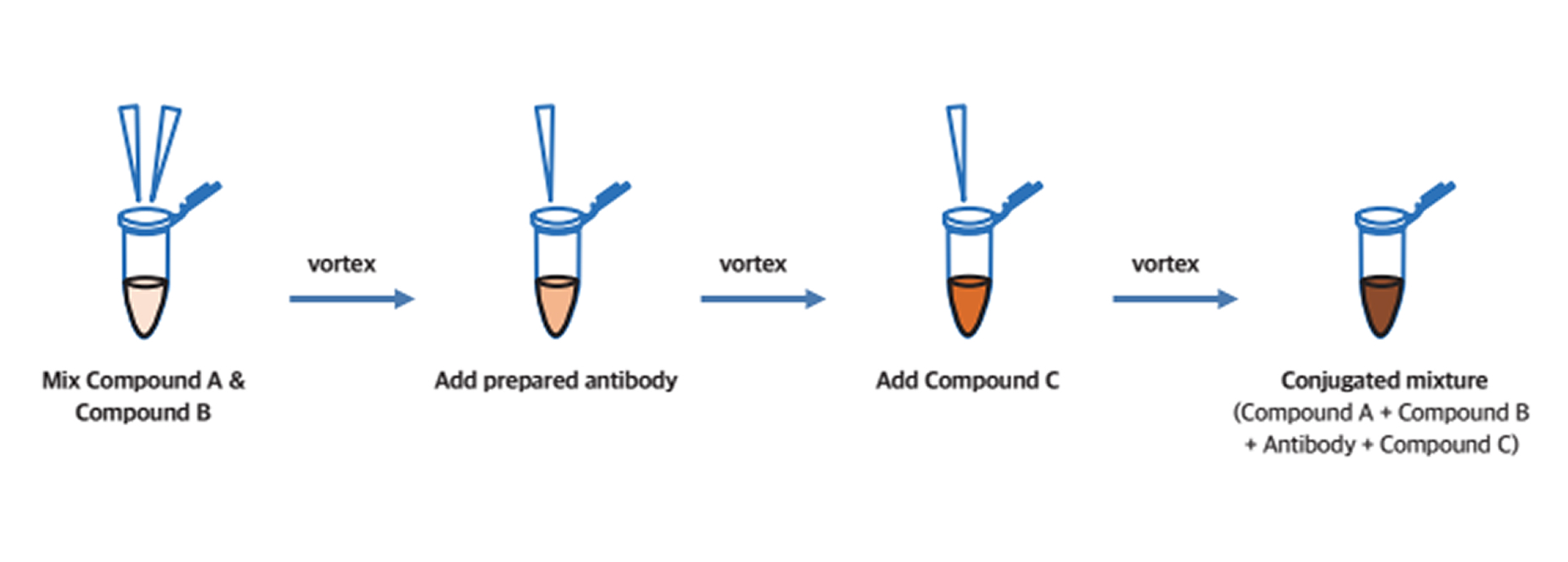
DIY In Vivo Labeling Kits
The IVI Tag™ DIY In Vivo Labeling Kit enables researchers to fluorescently tag their own targets—including antibodies, exosomes, peptides, and chemical drugs—for precise in vivo imaging. Using IVIM’s conjugation technology, the kit simplifies the labeling process into a few straightforward steps, making it possible to generate high-quality in vivo labels within hours. Unlike traditional antigen-antibody conjugation methods, IVI Tag™ DIY ensures that fluorophores are expressed at the intended target sites in living tissues, reducing variability and signal loss. This flexibility allows scientists to customize their experiments and track novel biological processes in real time.
In Vivo Label Preparation Steps
Conjugation
In Vivo Imaging Fixation Adjuncts
Stable, long-term imaging of dynamic organs with precision.
From brain to pancreas, expand research possibilities without compromising animal welfare.
Research Background
Intravital microscopy enables visualization of biological processes in living organisms, but motion artifacts, limited access, and repeated surgeries restrict long-term and reproducible imaging.
Fixation adjuncts solve these issues by stabilizing organs and providing optical access windows, allowing researchers to conduct weeks of continuous observation without sacrificing animals after each session.
Core Technologies
Organ-Specific Windows
Dorsal skinfold, cranial, abdominal, mammary, pancreas, and uterus imaging chambers designed for precise access.
Dynamic Organ Stabilization
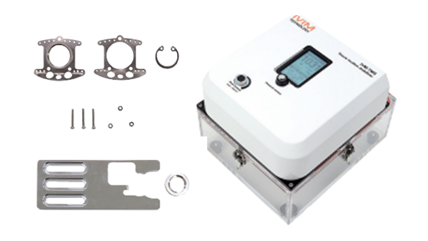
Tissue Motion Stabilizer (IVM-TMS) with micro-suction prevents micron-level tissue movement during imaging of the heart, lung, and thymus.
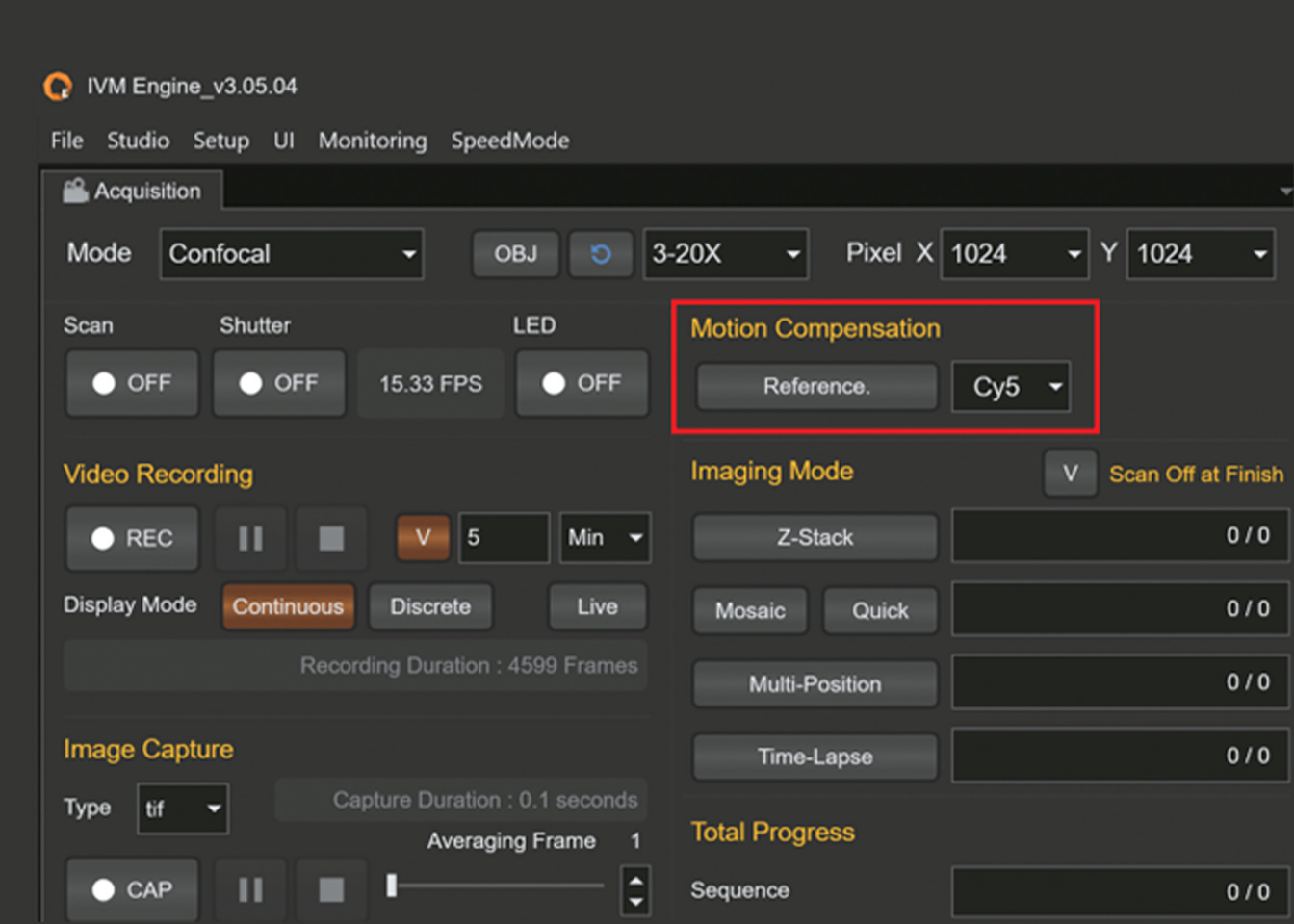
Motion Compensation Software
GPU-assisted algorithm corrects motion artifacts in real time, ensuring high-quality imaging of moving tissues.
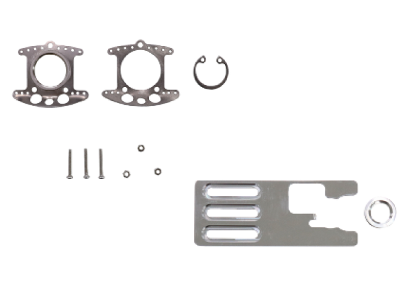
Minimally Invasive Design
Lightweight, compact chambers reduce surgical burden and extend longitudinal studies.
Research Benefits
- Enables longitudinal imaging for several weeks
- Reduces animal use, promoting ethical research practices
- Provides stable platforms for drug delivery, cancer progression, immune dynamics, and vascular studies
- Enhances reproducibility and quantitative reliability in preclinical models
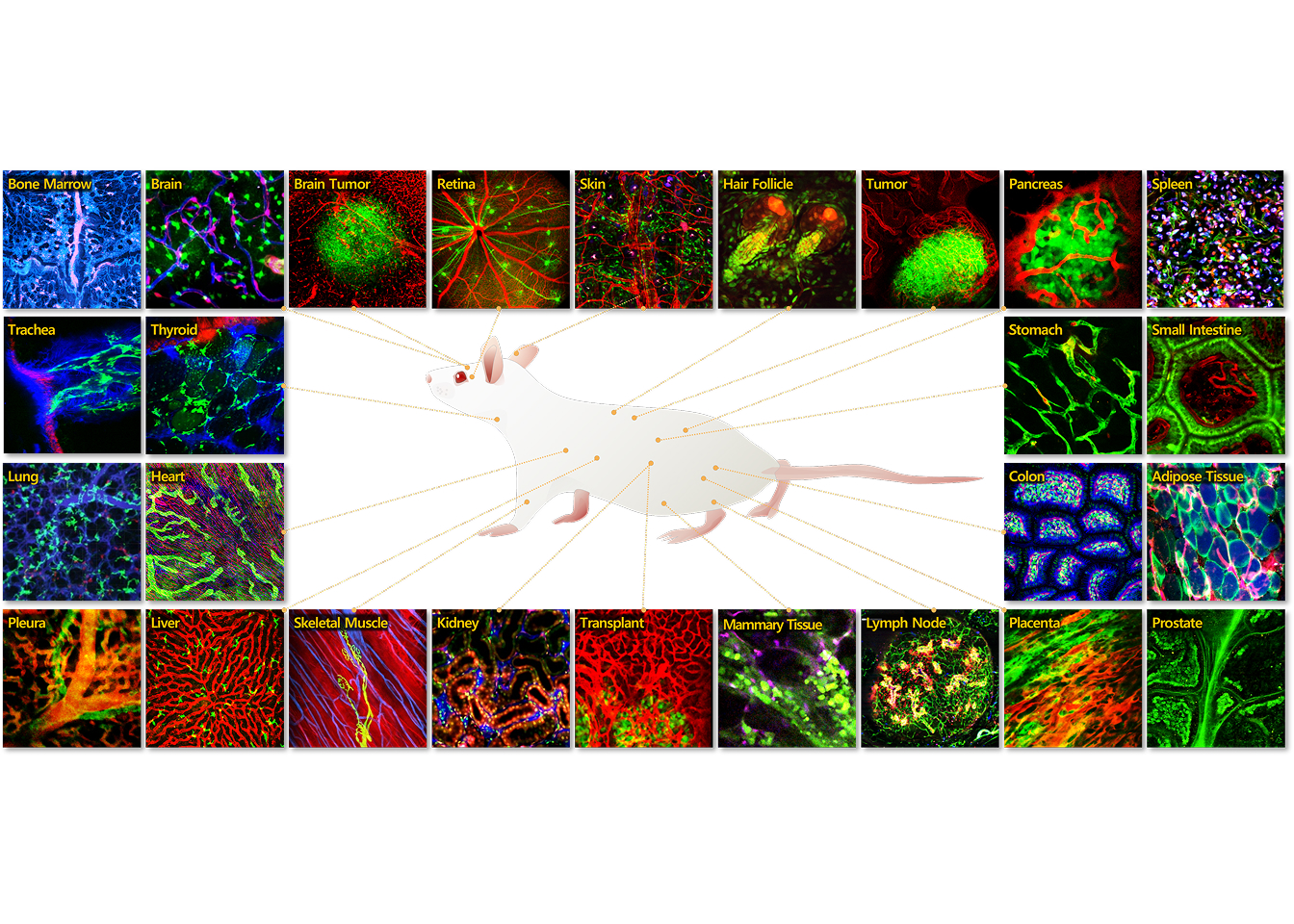
Applications Gallery
-
Lung
Intravital in vivo applications in the lungs of mice provide a powerful tool for studying pulmonary biology, disease processes.
-
Heart
In vivo intravital imaging of the heart in mice offers a powerful approach to studying dynamic cardiac processes and cardiovascular function.
-
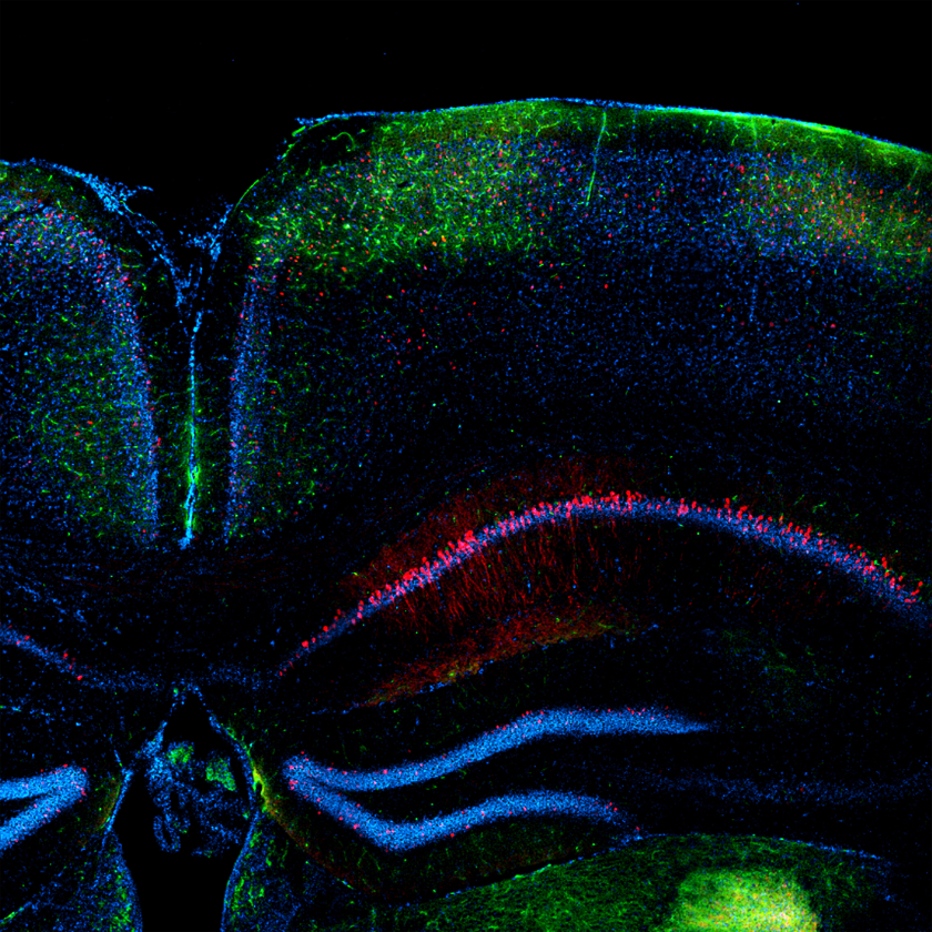
Brain
IVIM Technology provides advanced imaging solutions that support a wide range of intravital imaging applications in the brain.
-
Kidney
In vivo intravital imaging of the kidney offers a powerful approach to studying dynamic cellular and molecular processes within this vital organ.
-
Pancreas
Intravital in vivo applications in the pancreas offer a unique window into the dynamic processes underlying pancreatic physiology.
-
Liver
In vivo intravital imaging of the liver offers a powerful approach to studying dynamic cellular and molecular processes within this vital organ.
-
Tumor
In vivo intravital imaging of the tumor offers you the opportunity to explore various aspects of tumor biology through intravital imaging.
-
Skin
Leveraging intravital imaging techniques, IVIM Technology empowers researchers to gain valuable insights into the dynamic processes occurring within the skin microenvironment in vivo.