Introducing IVIM's In Vivo Labels for High-Quality IVIM TECHNOLOGY
NEWSLETTER
Volume 7, May 2025 |
|
|
About IVIM Technology, Inc.
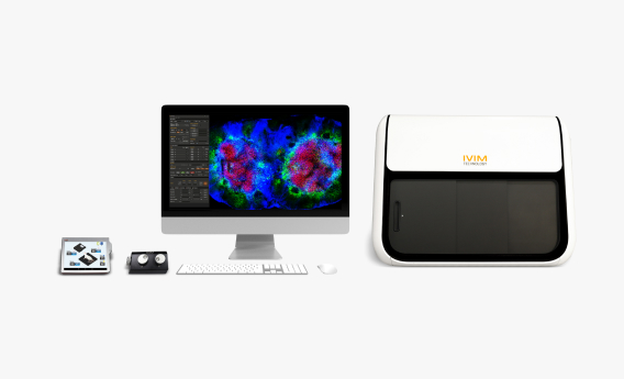
Founded in 2017, IVIM Technology is a leader in all-in-one turnkey intravital microscopy solutions. Our state-of-the-art, combined confocal and two-photon microscope systems provide real-time cellular imaging of live animals under anesthesia, revolutionizing in vivo research.
With a strong global presence and over 30 units sold, IVIM has earned recognition as one of the Top 10 APAC Bioanalytical Service Providers. We proudly serve over 100 organizations worldwide across academia, biotech, and the pharmaceutical industries. Our comprehensive range of solutions includes intravital microscopy (IVM), pre-clinical in vivo imaging, training, accessories, and labeled antibodies, enabling researchers to accelerate their scientific discoveries with precision and efficiency.
|
|
|
Elevating Intravital Imaging with AI:
Introducing the AI-Image Denoiser |
|
|
At IVIM Technology, we're always striving to push the boundaries of what's possible in high-resolution, real-time biological imaging. Intravital microscopy has already revolutionized the way researchers observe cellular behavior and drug distribution in living tissues. But with ultrafast imaging comes a well-known challenge: compromised image quality due to lower Signal-to-Noise Ratio (SNR).
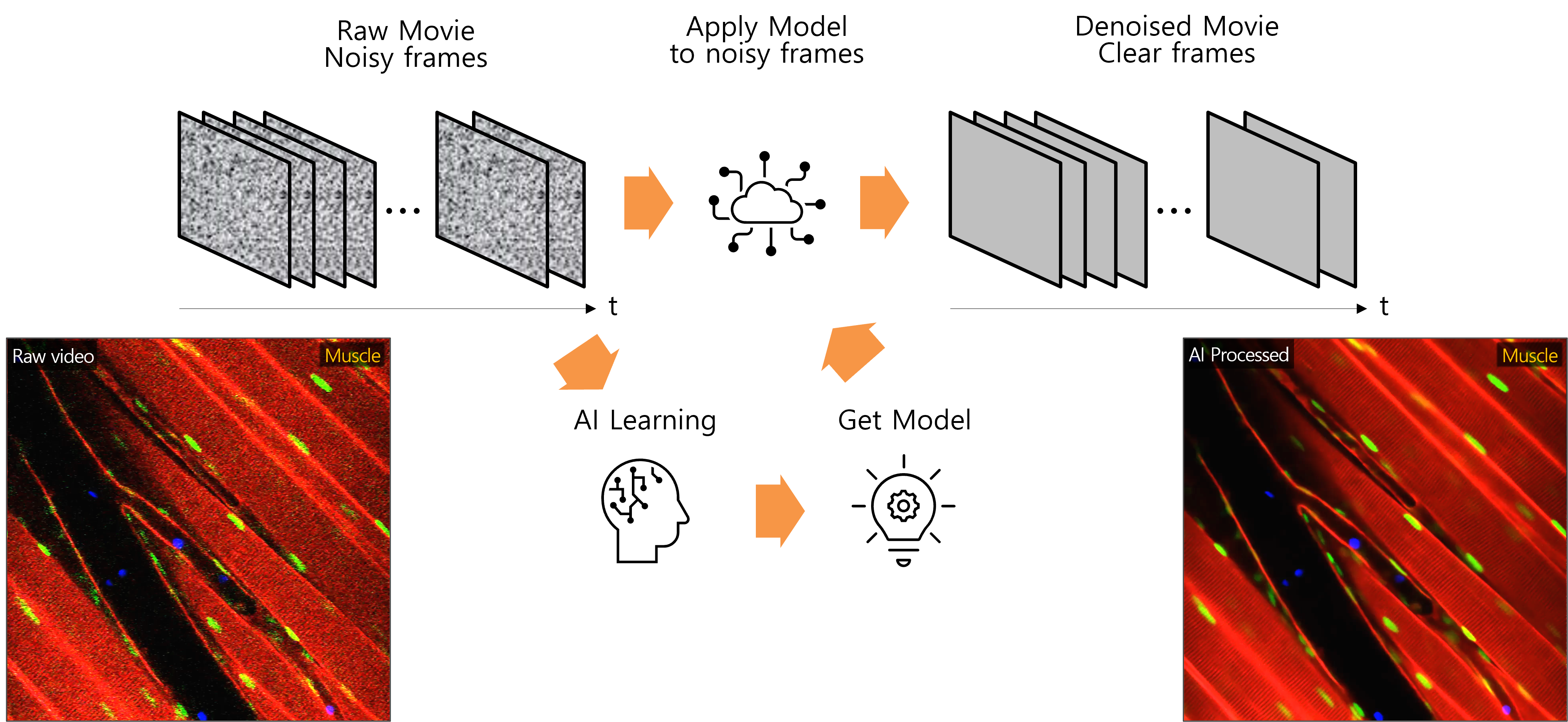
To observe this hurdle, we are proud to unveil AI-Image Denoiser, our best-in-class, AI-powered image enhancement software designed specifically for high-speed intravital microscopy. Officially launching on May 15, 2025, this self-supervised learning solution delivers remarkable improvements in image clarity and processing speed, all without requiring pre-trained data. |
|
|
Figure 1. Principle of AI-Image Denoiser |
|
|
Where traditional non-pretained AI tools may take over 7 hours to process large image sequences, AI-Image Denoiser completes the task in under 30 minutes, dramatically streamlining workflows and enabling real-time insight generation. More importantly, it intelligently distinguishes signal from noise to reveal previously obscured biological detail.
This new technology enables clearer, more accurate visualization of fast-moving live cells even in challenging, noisy imaging environment. By learning directly from the data, the AI-Image Denoiser intelligently enhances true signals while suppressing noise, offering a major leap forward in both the precision and efficiency of ultrafast intravital imaging. |
|
|
Figure 2. Cell counting analysis after AI denoising in intravital imaging of skin |
|
|
In a recent application using intravital microscopy on H2B-GFP transgenic mice, we observed skin tissue labeled to show blood vessels (red), blood flow (blue), and cell nuclei (green). Prior to denoising, the images were heavily affected by noise, resulting in blurred nuclei and poorly defined boundaries.
After applying the AI-Image Denoiser, the noise was significantly reduced, making the nuclear structures much clearer. This improvement allowed for accurate thresholding and cell counting using image analysing tool. While the raw images tended to overestimate cell numbers due to poor separation, the denoised results supported precise segmentation and reliable quantification. |
|
|
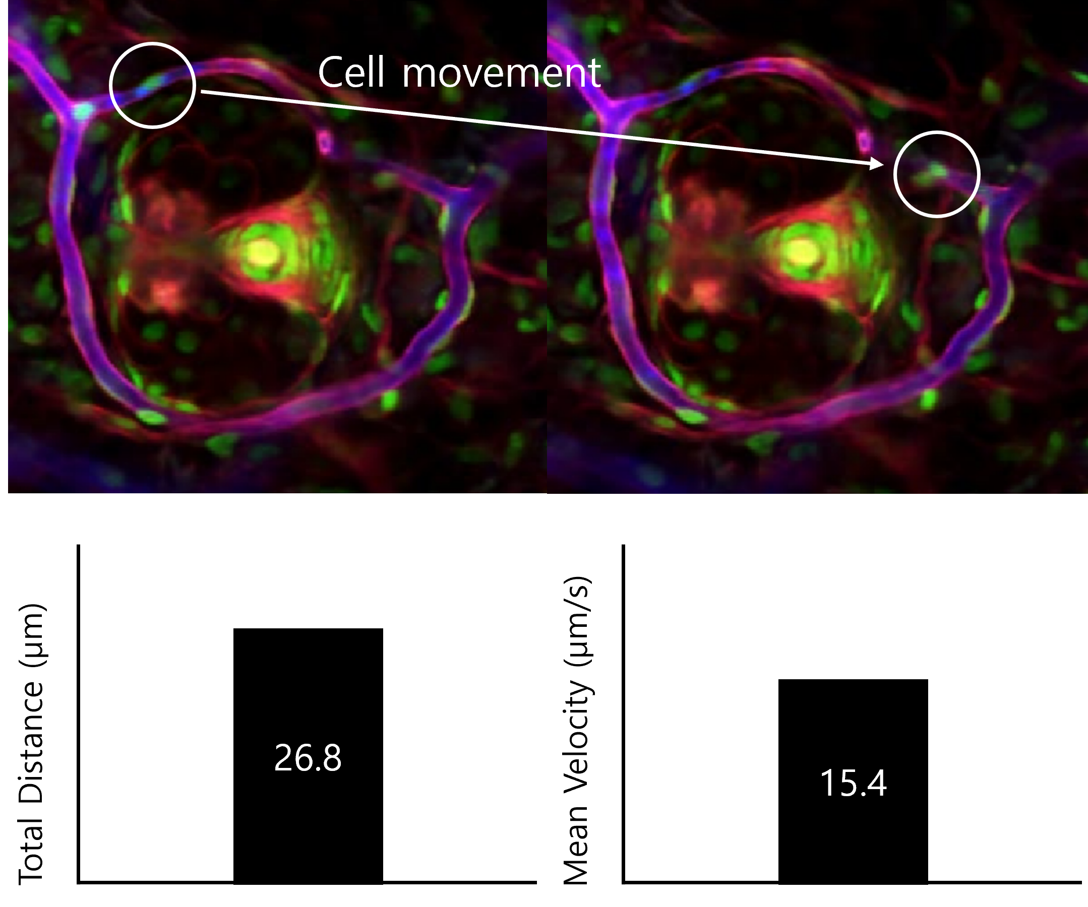
Figure 3. Cell tracking analysis after AI denoising in intravital imaging of skin |
|
|
Beyond cell counting, the denoiser also plays a key role in motion analysis. In previously noisy sequences where tracking was unreliable, AI-processed images revealed clear cell trajectories. Researchers can now analyze frame-by-frame cell movements, calculate displacement, and assess velocity with confidence.
By dramatically enhancing image clarity and reducing processing time, the AI-Image Denoiser transforms intravital microscopy into a more powerful tool for dynamic biological research. Whether you're investigating immune responses, tissue regeneration, or drug delivery, this innovation opens the door to deeper, more accurate insights, which delivered faster than before. |
|
|
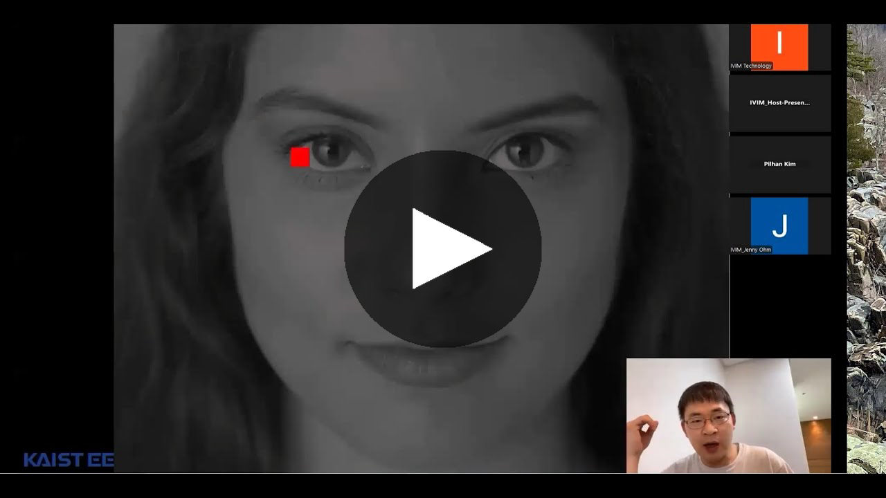
Watch Now! Webinar on AI-Powered Image Denoising for Intravital Imaging
The full session of the webinar is now available to watch on YouTube!
In this special webinar titled High-Resolution Intravital In Vivo Imaging with AI-Powered Image Denoising, experts Dr. Yong-Gyu Yoon and Dr. Pilhan Kim (Korea Advanced Institute of Science and Technology, KAIST) dive deep into how AI is transforming in vivo imaging. The session showcases IVIM Technology's All-in-One real-time intravital two-photon and confocal microscopy system, designed for cellular-level imaging of internal organs in live animal models of human diseases.
You'll also see a real-time demonstration of the AI-Image Denoiser software, which dramatically improves image clarity and processing efficiency for high-resolution intravital microscopy data.
Whether you're looking to streamline your imaging workflow or enhance the accuracy of your biological insights, this recorded session offers valuable takeaways on how AI is unlocking a new era of precision in intravital imaging.
Click below ↓↓↓ |
|
|
Upcoming Webinar Series
- JUN 26 | Evaluating the Efficacy of Antibody Therapeutic Drugs in Animal Models Using In Vivo Imaging
- JUL 23 | Advanced Imaging-Based Analysis of Neurobiological and Pharmacological Processes in Subcortical Brain Regions
- AUG 21 | From IVIS to IVM: Assessing the Efficacy of Small Molecule-Based Delivery Treatments with Intravital Microscopy
- AUG 28 | Real-Time In Vivo Imaging of Cellular Dynamics Using Advanced Intravital Microscopy
- SEP 24 | Cellular-Level In Vivo Mapping of Chemical Substance Distribution in Live Animals
- OCT 23 | Intravital Imaging of Blood Vessel Recovery and Angiogenesis for Development of Cell Therapeutics
- OCT 29 | Capturing Cellular Change in Real Time: The Power of Longitudinal In Vivo Imaging
And More... |
|
|
IVIM TECHNOLOGY. All Rights Reserved.
|
|
|
IVIM Technologyinformation@ivimtech.com#A-1305, Hyundai Knowledge Industry Center, 11, Beobwon-ro 11-gil, Songpa-gu, Seoul, 05836, Korea
Tel: +82-2-431-7450Unsubscribe |
|
|
|
|