In celebration of World Heart Day, explore 3 key disease IVIM TECHNOLOGY NEWSLETTER
Special Volume 11, September 2026 |
|
|
About IVIM Technology, Inc.
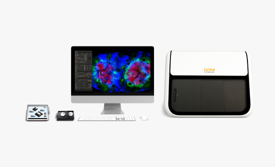
Founded in 2017, IVIM Technology is a global leader in turnkey intravital microscopy solutions. Our all-in-one box confocal and two-photon microscope systems enable real-time cellular imaging in live, anesthetized animals—transforming the landscape of in vivo research.
Recognized as one of the Top 10 APAC Bioanalytical Service Providers, IVIM serves a broad network of over 100 institutions worldwide, spanning academia, biotechnology, and the pharmaceutical industry. Our integrated portfolio includes intravital microscopy (IVM) systems, preclinical in vivo imaging solutions, training, specialized accessories, and labeled antibodies—empowering researchers to drive scientific discovery with precision, consistency, and efficiency.
|
|
|
World Heart Day:
Visualize the Injured Heart in Real Time |
|
|
September 29 marks World Heart Day, a global campaign to raise awareness of cardiovascular health and the importance of preventing heart disease.
The heart is more than a pump. It is a highly dynamic organ where blood continuously flows, immune cells migrate, and blood vessels and lymphatic vessels interact in real time.
But what actually happens inside the living heart when disease occurs?
Intravital imaging offers a unique window into these dynamic processes, enabling researchers to visualize blood flow, thrombus formation, immune cell responses, and changes in the cardiac microenvironment in real time.
This World Heart Day, we take a closer look at how the living heart responds to injury and metabolic stress through IVIM’s intravital imaging studies from acute myocardial infarction and microinfarction to high-fat diet-induced changes in the cardiac microenvironment. |
|
|
3 Key Disease Applications
for Cardiovascular Research |
|
|
01. When Blood Flow Stops
Visualizing Inflammation After Acute Myocardial Infarction
Acute myocardial infarction (AMI) causes rapid changes in the cardiac microenvironment, including impaired blood flow, inflammatory cell recruitment, and alterations in lymphatic function. Understanding these dynamic responses is important for investigating the progression of cardiac injury and subsequent tissue recovery.
Mouse model: Acute myocardial infarction (AMI) mouse model
1) Blood Flow & Neutrophil Recruitment
- Model & Imaging Approach
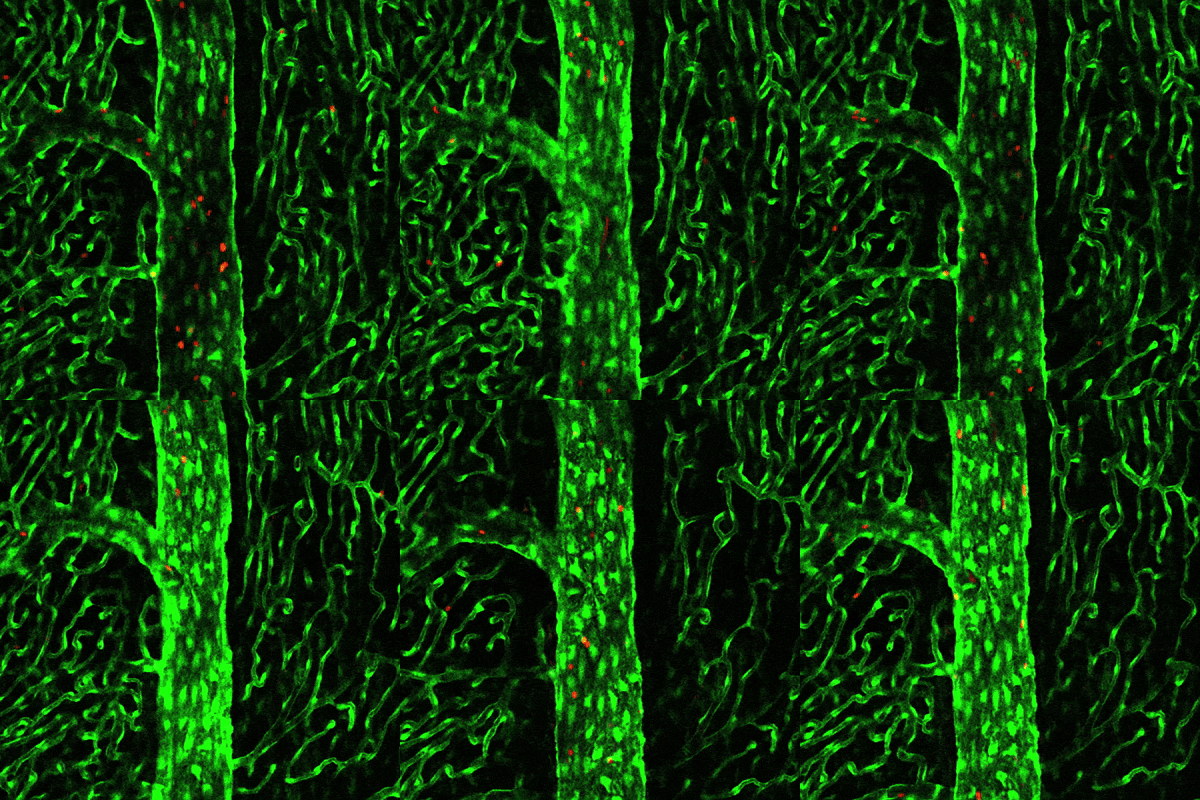
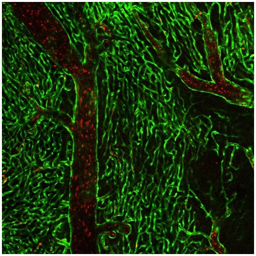
An AMI mouse model was established using a ligation-induced ischemia-reperfusion approach. Tie2-GFP transgenic mice were used to visualize cardiac blood vessels, while fluorescently labeled RBCs and Ly6G antibodies enabled real-time visualization of blood flow and neutrophil dynamics.
|
|
|
- Key Imaging Findings
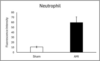
Compared with sham mice, AMI hearts showed markedly reduced or arrested blood flow together with increased neutrophil accumulation. The neutrophil signal was approximately 6-fold higher in the AMI group than in the sham group.
|
|
|
- Research Insight
These results demonstrate the ability of intravital imaging to simultaneously monitor vascular perfusion and immune cell recruitment following myocardial infarction, providing a dynamic view of the relationship between impaired blood flow and acute inflammation.
|
|
|
2) Lymphatic Vessel & Immune Response
- Model & Imaging Approach
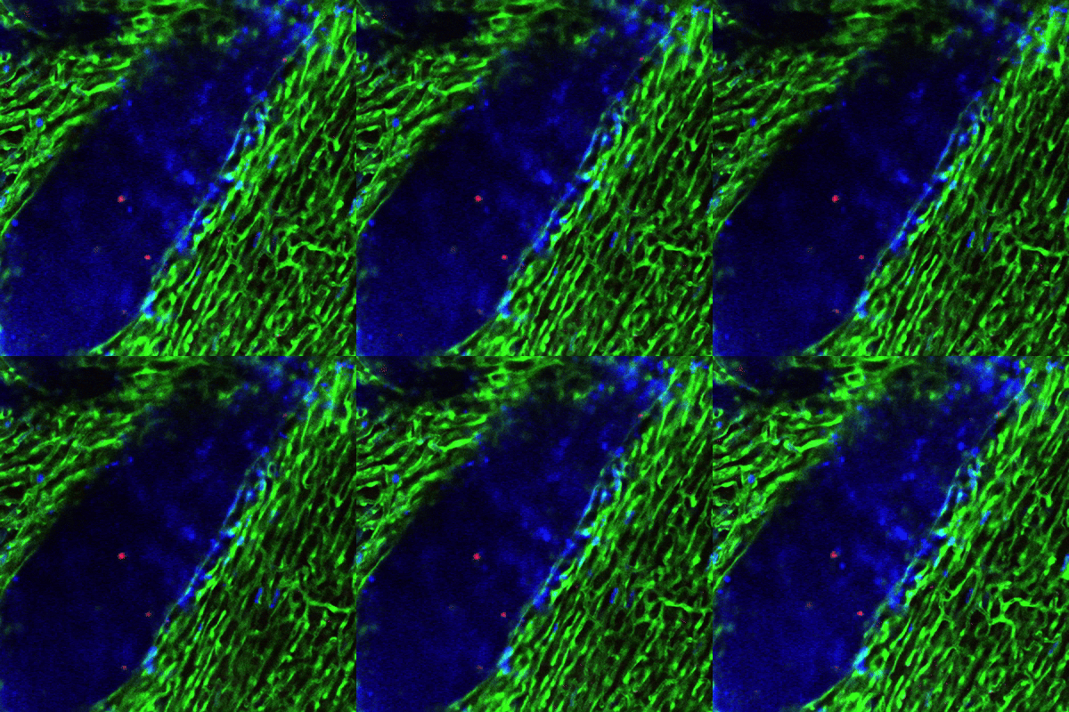
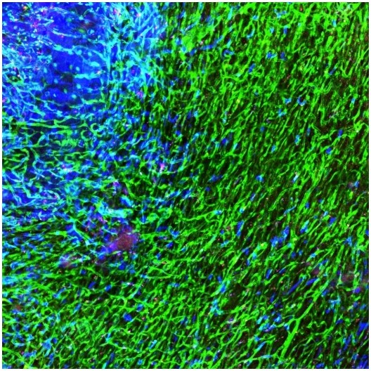
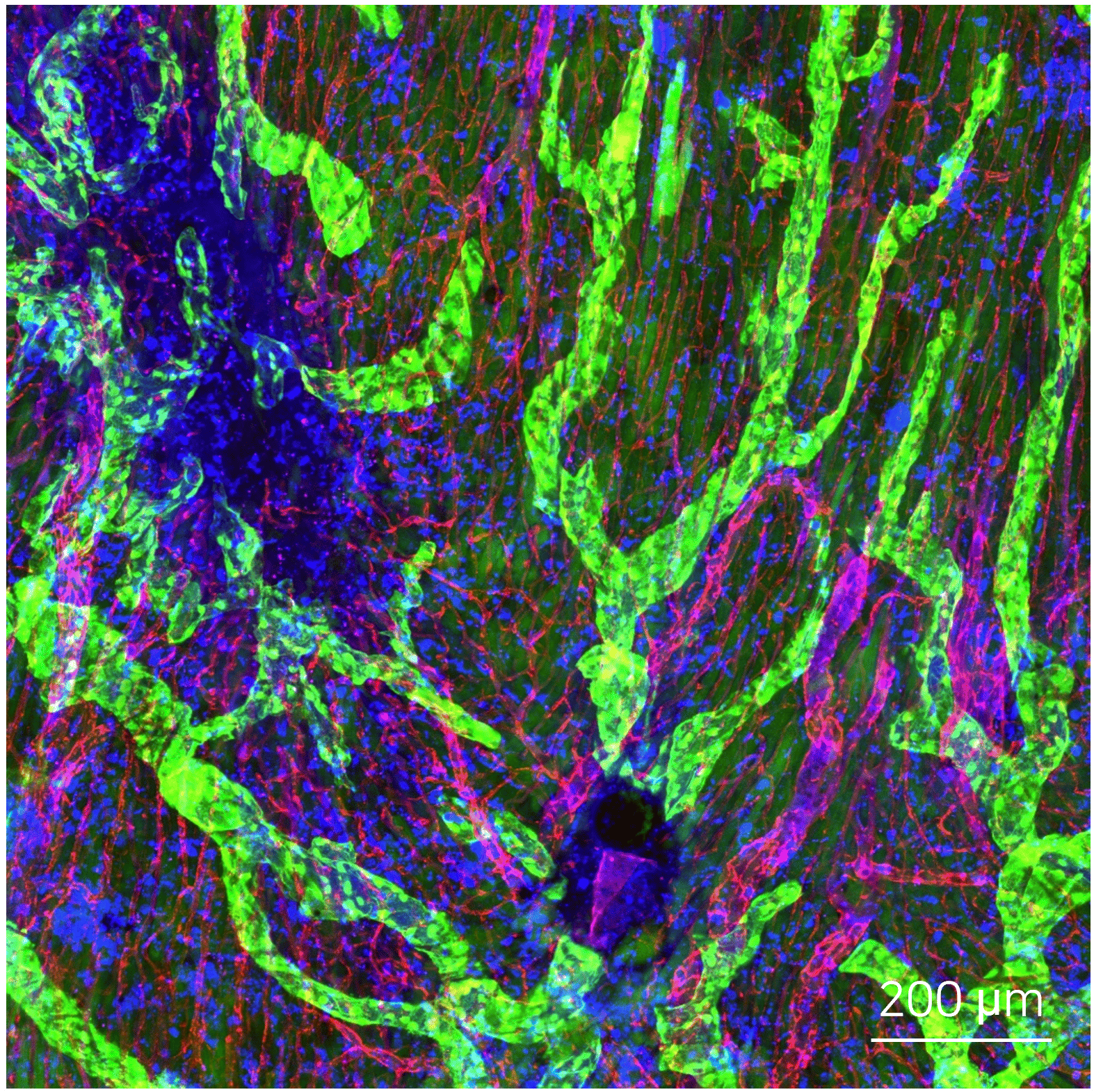
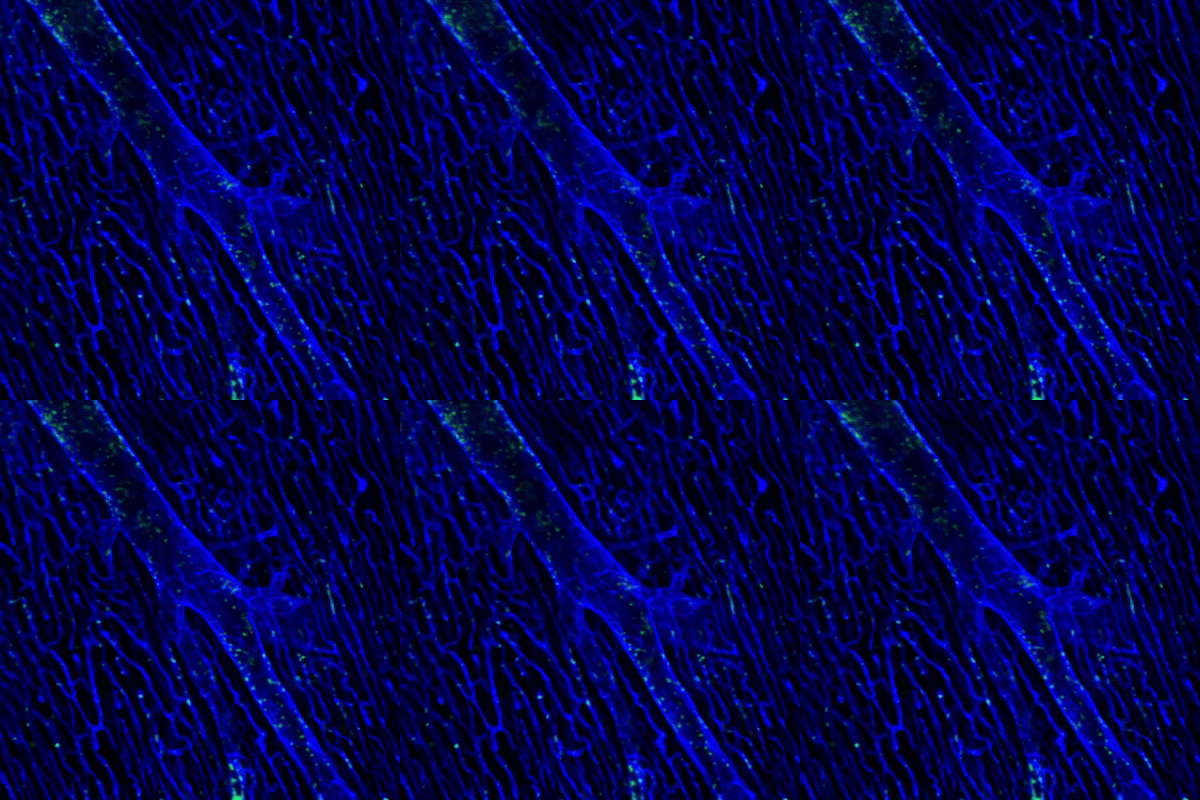
Prox1-GFP transgenic mice were used to visualize cardiac lymphatic vessels. Blood vessels and neutrophils were simultaneously labeled with CD31 and Ly6G, respectively, enabling the spatial relationship between lymphatic structures and immune cell accumulation to be examined in vivo.
- Key Imaging Findings
Compared with sham controls, AMI hearts showed increased neutrophil accumulation and thinner lymphatic vessels, particularly in regions with high neutrophil accumulation.
- Research Insight
The imaging results suggest a potential association between lymphatic alterations and neutrophil accumulation following myocardial infarction. This approach provides an opportunity to investigate how changes in cardiac lymphatic function may influence immune cell clearance and the inflammatory response after cardiac injury.
|
|
|
02. Watching a Thrombus Form
Real-Time Imaging of Myocardial Microinfarction
Myocardial microinfarction can result from localized thrombus formation and microvascular obstruction. Visualizing these events in real time is important for understanding how platelet accumulation and vascular occlusion contribute to cardiac tissue injury.
- Model & Imaging Approach
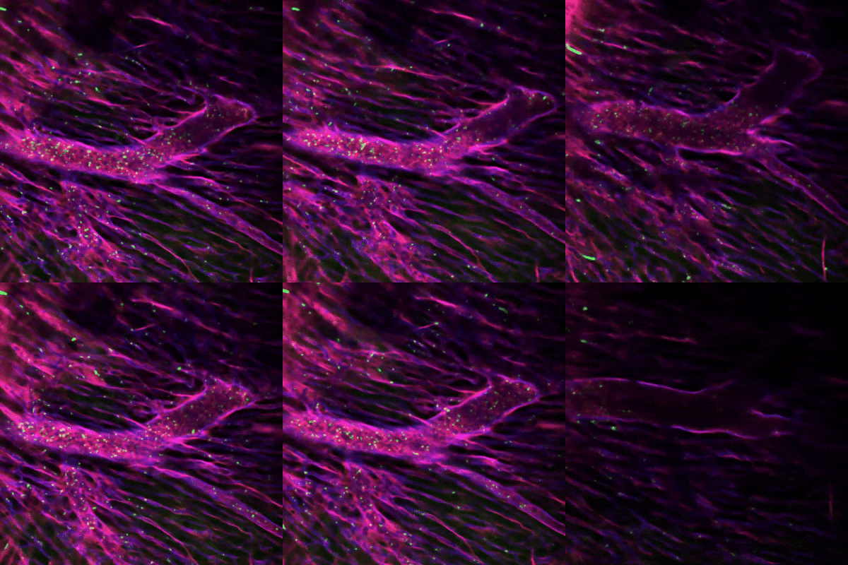
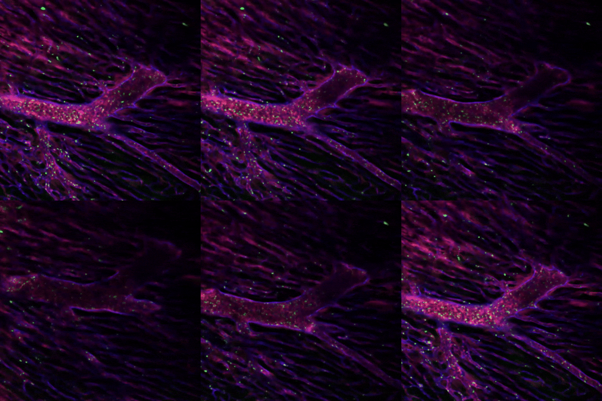
A Rose Bengal-mediated photothrombosis model was established in C57BL/6 wild-type mice. Following Rose Bengal administration, 561 nm laser illumination was used to induce localized thrombus formation. Blood vessels and platelets were visualized using CD31 and CD41 labeling, respectively. FSD555 was used as a fluorescent dye control to evaluate whether laser illumination alone induced changes in platelet accumulation or blood flow.
- Key Imaging Findings
Following laser illumination, progressive platelet accumulation was observed within cardiac blood vessels, accompanied by disruption and eventual arrest of blood flow. In contrast, the fluorescent dye control showed no noticeable changes in platelet accumulation or blood flow under the same illumination conditions.
- Research Insight
This model enables real-time visualization of the sequence of thrombus formation and blood flow disruption during myocardial microinfarction. It provides a useful platform for investigating platelet vascular interactions, microvascular obstruction, and the early events associated with ischemic cardiac injury.
|
|
|
03. Inside a Fat-Stressed Heart
Mapping Lipid Accumulation and Immune Cell Dynamics
Excessive lipid accumulation around the heart is associated with metabolic and inflammatory changes in the cardiac microenvironment. However, the spatial relationship between lipid deposition and immune cell responses in living tissue can be difficult to capture using conventional endpoint analyses.
- Model & Imaging Approach
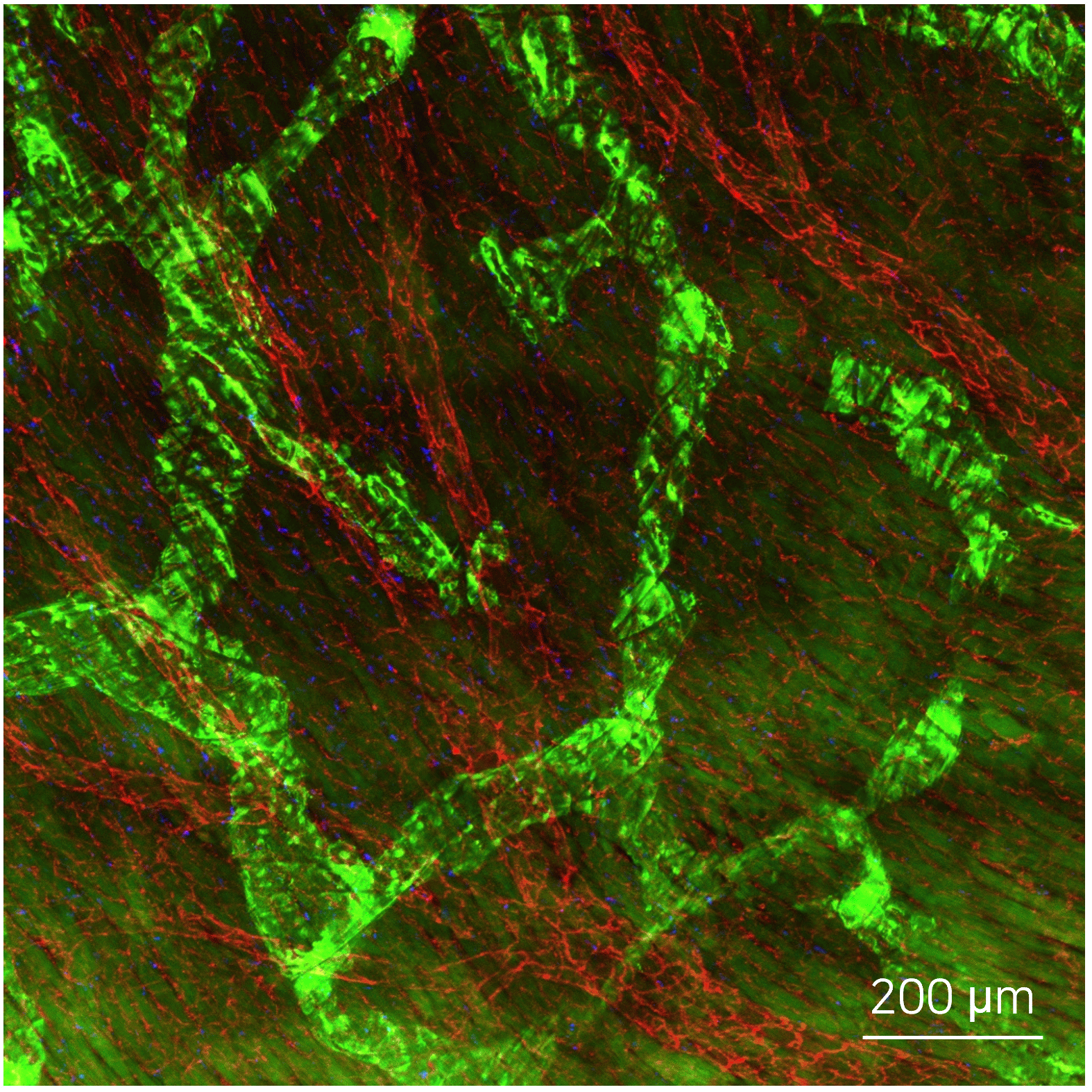
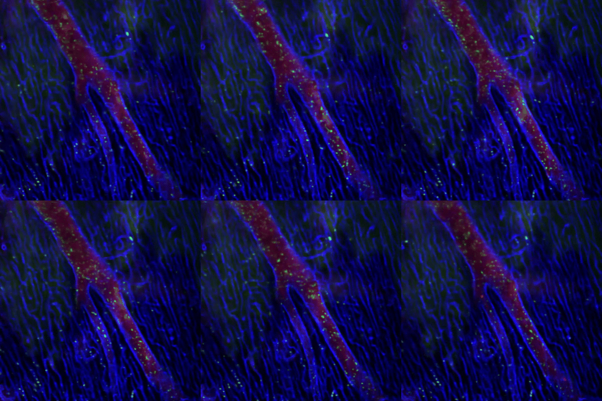
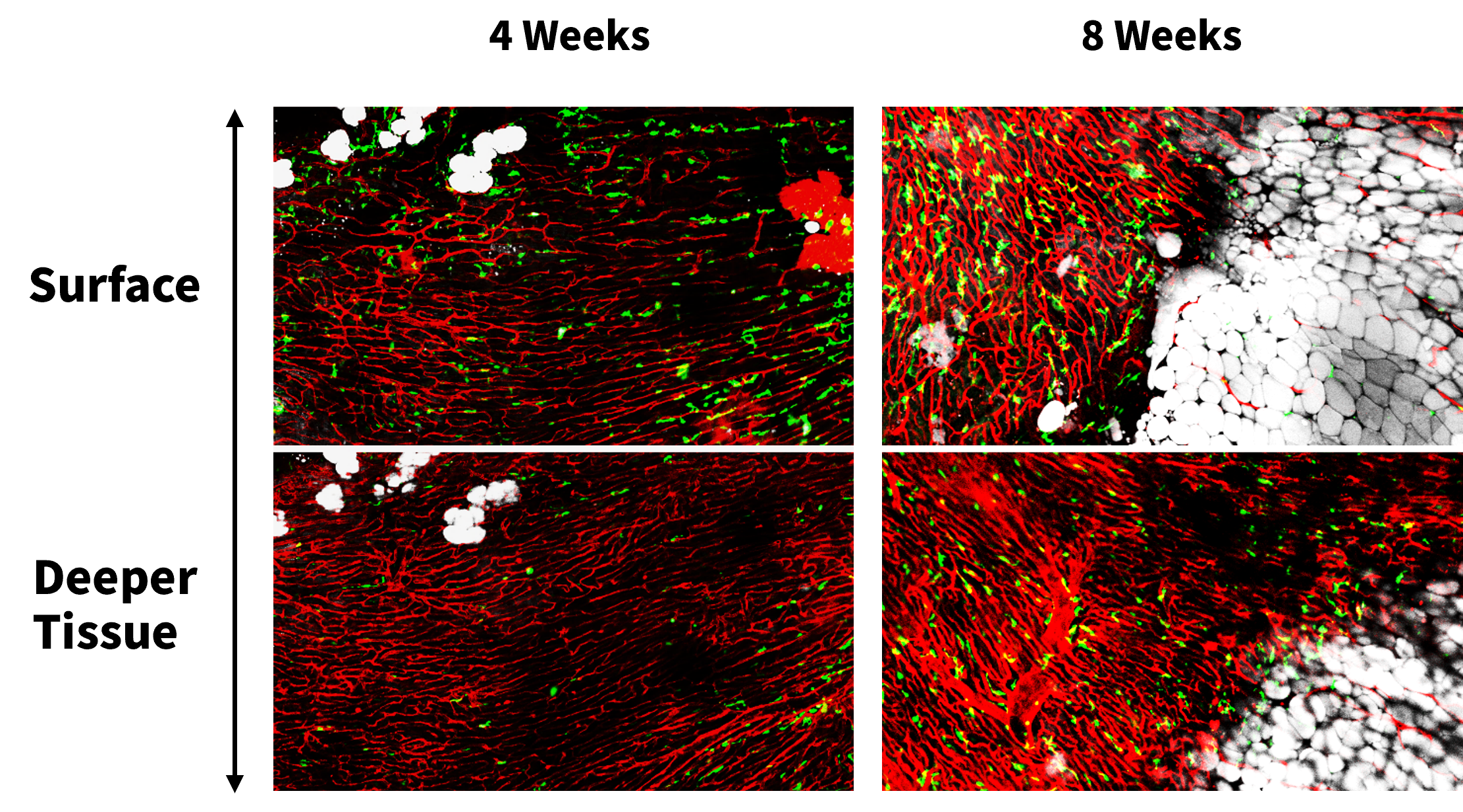
LysM-GFP transgenic mice were fed a high-fat diet for up to 8 weeks. Cardiac blood vessels were labeled with CD31, immune cells expressing LysM-GFP were visualized in vivo, and lipid droplets were labeled with SF44. Z-stack imaging was performed from the cardiac surface into deeper tissue layers to examine the spatial distribution of lipids and immune cells.
- Key Imaging Findings
Substantial lipid droplet accumulation was observed on the cardiac surface, while immune cells were predominantly detected in deeper tissue regions. Compared with the 4-week stage, 8 weeks of high-fat diet exposure resulted in increased lipid deposition accompanied by greater immune cell infiltration.
- Research Insight
These findings reveal distinct spatial patterns of lipid accumulation and immune cell localization within the heart. By imaging across multiple tissue depths, intravital microscopy provides a dynamic and spatially resolved view of how prolonged high-fat diet exposure alters the cardiac microenvironment and is associated with progressive immune responses.
|
|
|
Featured Paper: Intravital Heart Imaging |
|
|
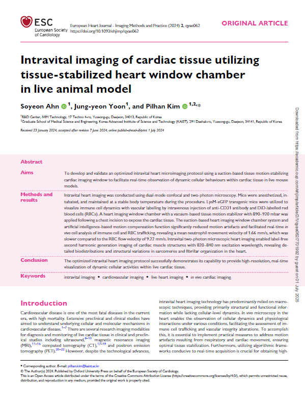
Explore a published study demonstrating how IVIM has been applied across heart research applications.
Title: Intravital imaging of cardiac tissue utilizing tissue-stabilized heart window chamber in live animal model
Journal: European Heart Journal |
|
|
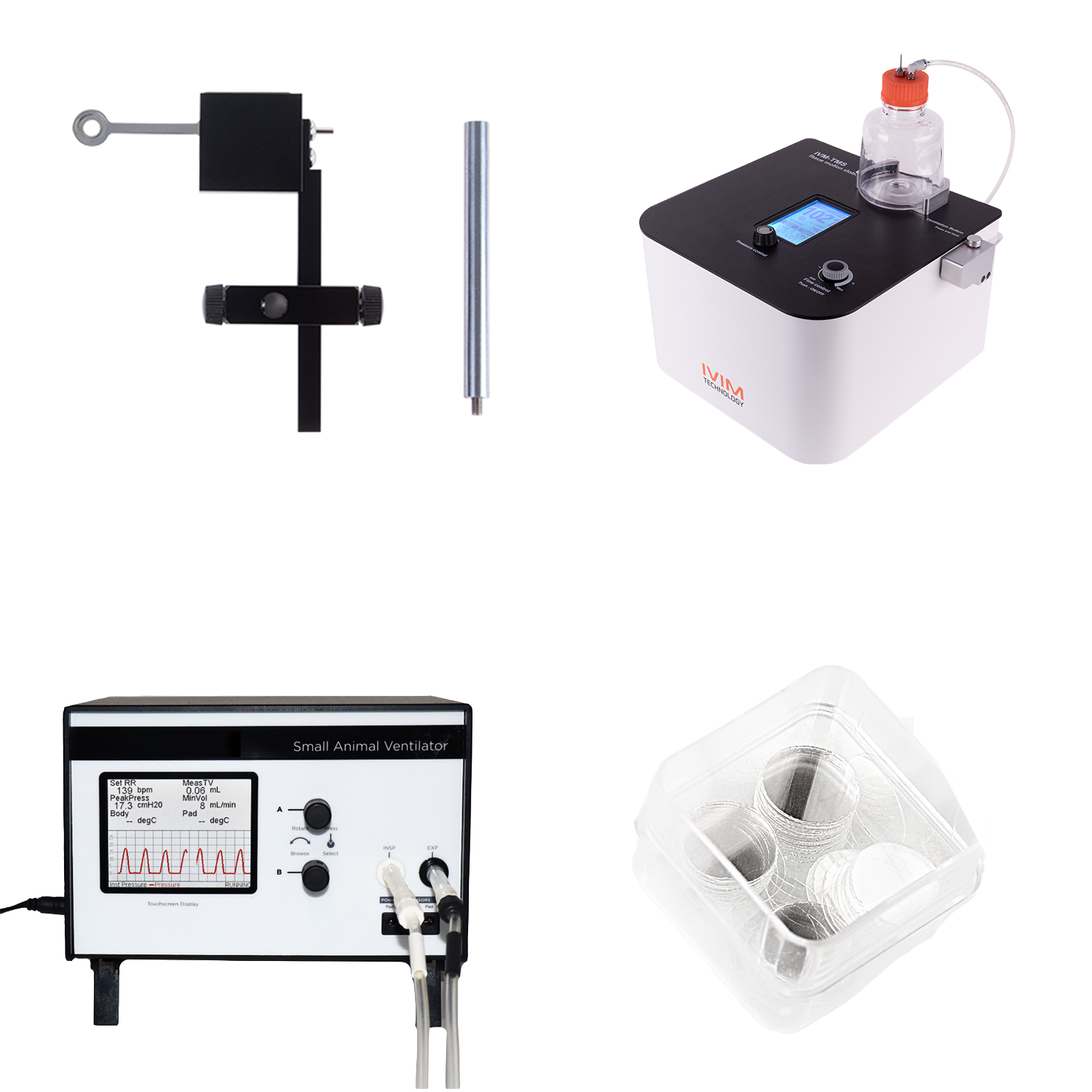
Curious about the accessories used in this study? |
|
|
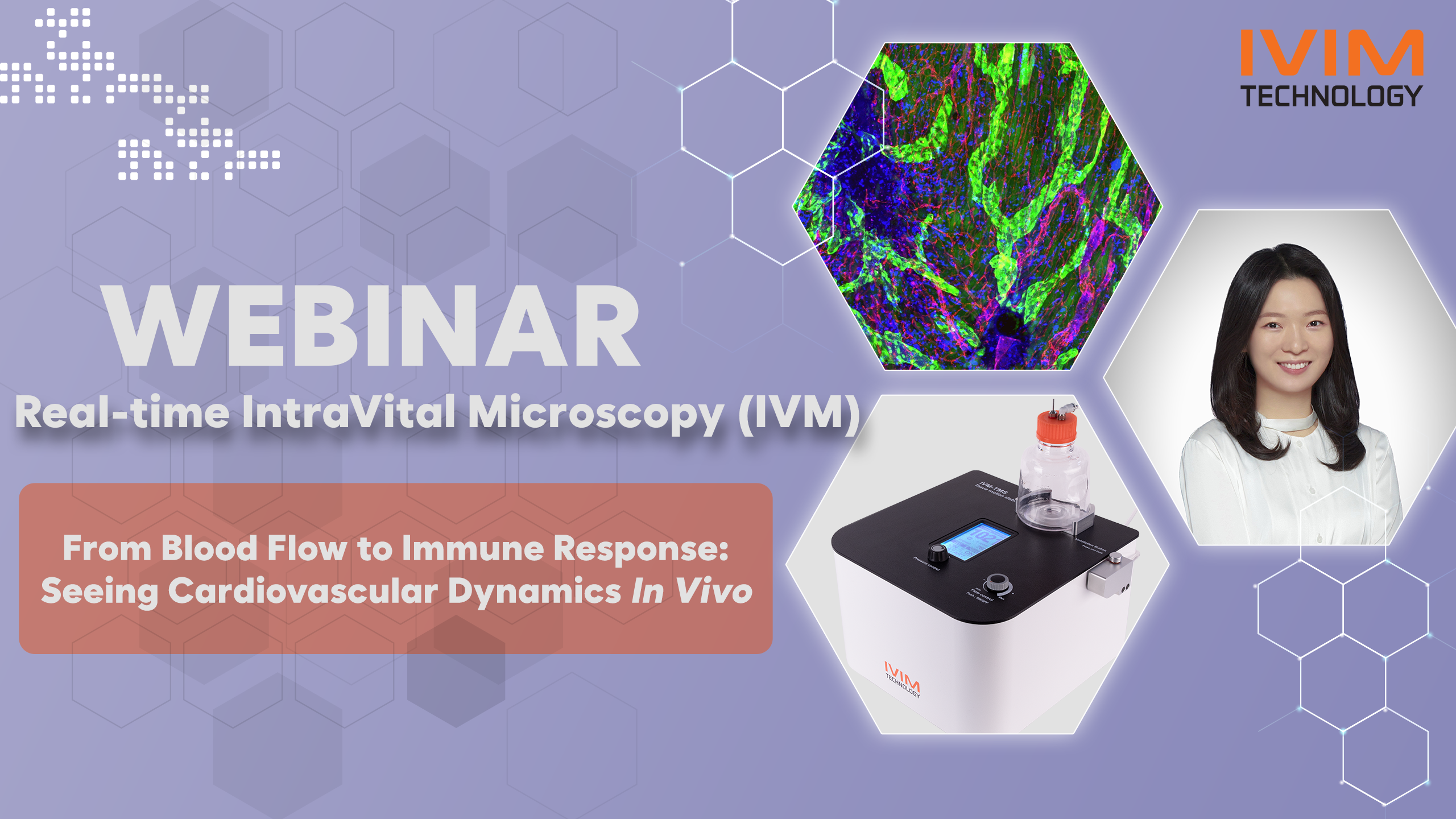
Explore the IVM-TMS (Tissue Motion Stabilizer) and Vacuum-Based Chamber, part of our optimized solution for intravital heart imaging! |
|
|
Upcoming Cardiovascualr Webinar |
|
|
DATE: October 22, 2026
TIME: 10:00 AM (EDT) / 9:00 AM (CDT) / 3:00 PM (WEST) / 4:00 PM (CEST)
|
|
|
Upcoming Vascular Exhibitions in Q3-Q4 2026
- October 2-3 | KVBM 2026 (Korean Society for Vascular Biology and Medicine)
- October 26-29 | SHVM 2026 (Society for Heart and Vascular Metabolism)
- November 14-15 | JVBMO 2026 (The 34th Annual Meeting of the Japanese Vascular Biology and Medicine Organization)
|
|
|
IVIM TECHNOLOGY. All Rights Reserved.
|
|
|
IVIM Technologyinformation@ivimtech.com#A-1305, Hyundai Knowledge Industry Center, 11, Beobwon-ro 11-gil, Songpa-gu, Seoul, 05836, Korea
Tel: +82-2-431-7450Unsubscribe |
|
|
|
|