Intravital in vivo cellular level animal imaging - Tumor
High-Resolution Intravital In Vivo Imaging
with AI-Powered Image Denoising
|
|
DATE: Thursday, May 15, 2025
TIME: 7:00 am PT; 10:00 am. ET; 4:00 pm. CET
DURATION: 60 mins
In this webinar, experts discuss cutting-edge advancements in microscopy imaging and denoising technologies.
SUPPORT: A Self-Supervised Learning Framework for Robust Denoising of Microscopy Images with Fast Dynamics
Young-Gyu Yoon, PhD, KAIST
Microscopy imaging plays a crucial role in modern biological research, but the inherent trade-offs between imaging speed and signal-to-noise ratio present significant challenges. As researchers push toward capturing faster biological processes, noise becomes increasingly problematic, potentially obscuring the very dynamics they aim to study. In this talk, Dr. Yoon presents SUPPORT (Statistically Unbiased Prediction Utilizing Spatiotemporal information in imaging data), a novel self-supervised learning method for removing Poisson-Gaussian noise in microscopy images. Unlike previous approaches that rely heavily on temporal redundancy, SUPPORT captures the spatiotemporal dependencies between neighboring pixels even when temporally adjacent frames provide limited information – making it particularly effective for imaging fast dynamic processes. He demonstrates how SUPPORT successfully denoises various microscopy datasets, including time-lapse fluorescence microscopy of freely moving organisms, volumetric structural imaging, calcium imaging, and voltage imaging while preserving the underlying biological dynamics. The method’s effectiveness across diverse imaging modalities suggests broad applicability in microscopy-based research.
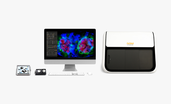
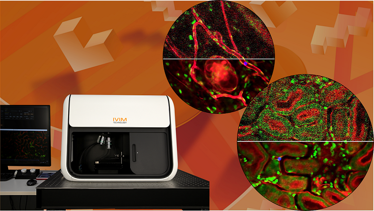
In Vivo Cellular-Level Imaging of Internal Organs in Live Animals Using AI-Image Denoiser Software
Pilhan Kim, PhD, Professor & CEO/CTO, Korea Advanced Institute of Science and Technology (KAIST) & IVIM Technology Inc.
Microscopy imaging is essential in biological research, but balancing speed and signal-to-noise ratio remains a significant challenge. In this webinar, we will introduce IVIM Technology’s All-in-One real-time intravital two-photon and confocal microscopy system, optimized for in vivo cellular-level imaging of internal organs in live animal models of human diseases. This advanced system can capture real-time, multi-color, sub-micron resolution images with an automatic motion compensation function, enabling the analysis of complex in vivo environments, including stromal cells, immune cells, vascular cells, and the extracellular matrix.
A key feature of the system is its integration of AI-powered imaging denoising, which enhances image quality by reducing noise and preserving cellular details. This technology allows for clearer visualization of dynamic cellular interactions in live tissue, even in noisy environments or when imaging fast-moving processes. Dr. Pilhan Kim will also discuss the system’s potential for capturing intricate cellular dynamics in real-time. Additionally, we will demonstrate in real-time how IVIM Technology’s AI-Image Denoiser Software effectively processes high-resolution intravital in vivo images.
Key Topics Include:
- Understand the Challenges of Microscopy Imaging
- Gain Insight into SUPPORT Methodology
- Understand the Broader Applicability of SUPPORT in Microscopy-Based Research
- Learn How AI-Powered Denoising Enhances Image Quality
- Discover the Applications of Real-Time Cellular Imaging
- See the Real-Time Demonstration of AI-Image Denoiser Software
|
IVIM Technology
information@ivimtech.com
#A-1305, Hyundai Knowledge Industry Center, 11,
Beobwon-ro 11-gil, Songpa-gu, Seoul, 05836, KoreaUnsubscribe |
|
|